 뒤로
Anatomy & Physiology: Nervous System Fundamentals
 03:35

이 집합의 용어 (33)
The nervous system has three overlapping functions: sensory input (monitoring changes), integration (processing and interpreting input), and motor output (activating effectors like muscles and glands).
The CNS consists of the brain and spinal cord, serving as the integrating and control center that interprets sensory input and dictates motor output.
The PNS includes all nervous tissue outside the CNS, mainly nerves and ganglia, linking the body to the CNS.
Consists of nerve fibers that carry impulses toward the CNS from sensory receptors, including somatic sensory fibers (from skin, muscles, joints) and visceral sensory fibers (from organs).
Transmits impulses away from the CNS to effector organs (muscles and glands), divided into the somatic nervous system (voluntary control of skeletal muscles) and autonomic nervous system (involuntary control of smooth muscle, cardiac muscle, glands).
Supporting cells in nervous tissue that surround and protect neurons; include astrocytes, microglia, ependymal cells, oligodendrocytes in CNS, and Schwann and satellite cells in PNS.
Most abundant CNS glial cells; support neurons, regulate chemical environment, guide neuron migration, and help form synapses.
Small CNS glia that monitor neuron health and transform into macrophages to phagocytize debris and pathogens.
CNS glial cells that wrap axons with myelin sheaths, insulating nerve fibers and increasing impulse speed.
PNS glial cells that form myelin sheaths around peripheral nerve fibers and aid in nerve regeneration.
Neurons are excitable, have extreme longevity, are amitotic (mostly non-dividing), and have a high metabolic rate requiring continuous oxygen and glucose.
Consists of a cell body (soma) with nucleus and organelles, dendrites (input regions), and a single axon (conducting region).
Bidirectional movement of substances along the axon: anterograde (away from cell body) and retrograde (toward cell body), using motor proteins kinesin and dynein.
Myelin electrically insulates axons and increases the speed of nerve impulse conduction.
In the PNS, Schwann cells form myelin sheaths; in the CNS, oligodendrocytes form myelin sheaths around multiple axons.
Neurons are classified as multipolar (most common), bipolar (two processes), and unipolar (single short process that splits).
Neurons are sensory (afferent), motor (efferent), or interneurons (association neurons within CNS).
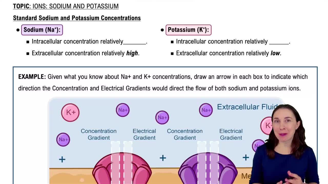
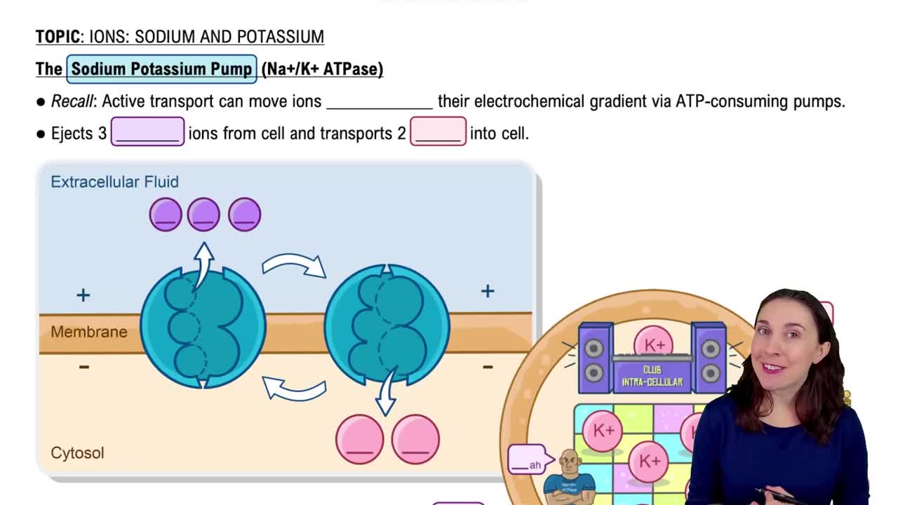
Typically about −70 mV in neurons, caused by differences in ion concentrations and membrane permeability, especially K+ and Na+ gradients.
Current (I) = Voltage (V) / Resistance (R); voltage is potential energy from separated charges, current is ion flow, and resistance is membrane hindrance.
Include chemically gated (ligand-gated), voltage-gated, and mechanically gated channels that open in response to specific stimuli.
Depolarization makes the membrane potential less negative (closer to zero or positive), increasing chance of firing; hyperpolarization makes it more negative, decreasing firing probability.
Short-lived, localized changes in membrane potential that vary in strength and decrease with distance; occur in dendrites and cell bodies.
Brief, long-distance signals in axons with a fixed amplitude (~100 mV), generated by voltage-gated Na+ and K+ channels, and do not decay with distance.
Include resting state, depolarization (Na+ channels open), repolarization (Na+ channels inactivate, K+ channels open), and hyperpolarization (K+ channels close slowly).
An action potential either occurs fully if threshold is reached or not at all if threshold is not reached.
APs are regenerated along the axon by local currents depolarizing adjacent membrane areas, moving in one direction away from the origin.
In myelinated axons, APs jump between nodes of Ranvier, increasing conduction speed about 30 times compared to continuous conduction in unmyelinated axons.
A junction where information is transmitted from one neuron to another or to an effector cell; includes presynaptic neuron, synaptic cleft, and postsynaptic neuron.
AP arrives → voltage-gated Ca2+ channels open → Ca2+ influx triggers neurotransmitter release → neurotransmitter diffuses and binds receptors → ion channels open → graded potentials generated.
Depolarizing graded potentials caused by neurotransmitter opening channels allowing Na+ influx > K+ efflux, increasing likelihood of AP generation.
Hyperpolarizing graded potentials caused by neurotransmitter opening K+ or Cl− channels, making the membrane more negative and less likely to fire an AP.
EPSPs and IPSPs combine spatially and temporally at the axon hillock to determine if threshold is reached and an AP is generated.
Repeated use of a synapse increases neurotransmitter release and postsynaptic response, enhancing synaptic efficiency and contributing to learning and memory.