Skip to main content
Anatomy & Physiology
나의 코스
배우기
시험 준비
AI 튜터
학습 가이드
교과서적 해결책
플래시카드
탐험해 보세요
앱을 사용해 보세요
나의 코스
배우기
시험 준비
AI 튜터
학습 가이드
교과서적 해결책
플래시카드
탐험해 보세요
앱을 사용해 보세요
뒤로
Anatomy & Physiology: Skeletal and Nervous System Key Concepts
카드를 뒤집기 위해 탭할 수 있습니다.
Identify cervical vertebrae
카드를 뒤집기 위해 탭할 수 있습니다.
👆
Identify cervical vertebrae
Cervical vertebrae have
transverse foramina
that allow passage of vertebral arteries.
진행 상황 추적
컨트롤 버튼이 '내비게이션' 모드로 변경되었습니다.
1/30
추천 영상
VideoThumbView.guidedCourse
6:29
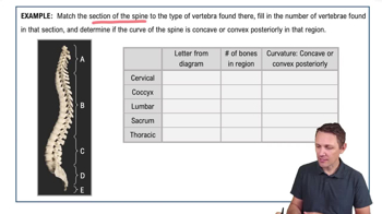
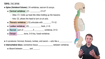
Spine (Vertebral Column)
12720
views
271
rank
VideoThumbView.guidedCourse
5:17
The Spine Example 1
9080
views
190
rank
이 집합의 용어 (30)
하이드의 정의
Identify cervical vertebrae
Cervical vertebrae have
transverse foramina
that allow passage of vertebral arteries.
Atlas and axis features
The first cervical vertebra, the
atlas
, lacks a body; the second, the
axis
, has a superior projection called the
dens (odontoid process)
.
Thoracic vertebrae identification
Thoracic vertebrae have long spinous processes and
costal facets
that articulate with ribs.
Lumbar vertebrae identification
Lumbar vertebrae have thick bodies and short, blunt spinous processes.
Intervertebral disc composition
An intervertebral disc has an outer fibrous ring called the
anulus fibrosus
and an inner gelatinous core called the
nucleus pulposus
.
Function of intervertebral discs
Intervertebral discs provide
shock absorption
and allow flexibility in the vertebral column.
Types of vertebral fractures
Wedge fracture
,
burst fracture
, and
herniated disc
differ in cause and symptoms, often involving trauma or disc displacement.
Purpose of a spinal tap (lumbar puncture)
A spinal tap collects cerebrospinal fluid for diagnosis or delivers medication; performed below the spinal cord end.
Structures of the thoracic cage
The thoracic cage includes the
sternum
,
ribs
, and
thoracic vertebrae
.
Function of the thoracic cage
It protects vital organs and supports respiration.
Correct CPR compression location
CPR compressions should be done on the
lower half of the sternum
to avoid injury.
Membrane potential definition
Membrane potential is the
voltage difference
across a cell membrane due to ion distribution.
Three main types of ion channels
Leak channels
,
gated channels
, and
voltage-gated channels
differ by how they open and close.
Resting membrane ion distribution
At rest,
Na+
is higher outside,
K+
is higher inside; gradients create electrical and chemical forces.
Cause of negative resting membrane potential
Movement of positive ions out of the cell (mainly K+) makes the inside more negative.
Neuron structure and function
Dendrites receive signals, the soma integrates, the axon transmits action potentials, and synaptic terminals release neurotransmitters.
Depolarization vs repolarization vs hyperpolarization
Depolarization: membrane becomes less negative; repolarization: returns to resting; hyperpolarization: more negative than resting.
Phases of an action potential
1) Depolarization: Na+ influx; 2) Repolarization: K+ efflux; 3) Hyperpolarization: continued K+ outflow.
Local potentials vs action potentials
Local potentials are graded and reversible; action potentials are all-or-none and propagate long distances.
Absolute vs relative refractory periods
Absolute: no new AP possible; relative: larger stimulus needed to trigger AP.
Saltatory conduction
In myelinated neurons, action potentials jump between nodes of Ranvier, increasing conduction velocity.
Chemical synapse transmission steps
Action potential triggers Ca2+ influx, vesicle fusion, neurotransmitter release, receptor binding, and postsynaptic response.
Excitatory vs inhibitory postsynaptic potentials
EPSPs depolarize and increase AP likelihood; IPSPs hyperpolarize and decrease AP likelihood.
Summation in neurons
Spatial and temporal summation combine local potentials to reach threshold for an action potential.
Bone organic and inorganic matrix roles
Organic matrix (collagen) provides flexibility; inorganic matrix (hydroxyapatite) provides strength.
Osteoblast role in intramembranous ossification
Osteoblasts produce bone matrix directly from mesenchyme.
Bone collar in endochondral ossification
The bone collar forms around the cartilage model, initiating ossification.
Cells responsible for longitudinal bone growth
Chondrocytes
in the epiphyseal plate divide to lengthen bones.
Appositional bone growth
Bone grows in thickness by osteoblasts adding matrix beneath the periosteum.
Bone remodeling in healthy bone
Bone remodeling continuously replaces old bone to maintain strength and mineral balance.
 뒤로뒤로
 6:29