Skip to main content
Anatomy & Physiology
나의 코스
배우기
시험 준비
AI 튜터
학습 가이드
교과서적 해결책
플래시카드
탐험해 보세요
앱을 사용해 보세요
나의 코스
배우기
시험 준비
AI 튜터
학습 가이드
교과서적 해결책
플래시카드
탐험해 보세요
앱을 사용해 보세요
뒤로
Anatomy & Physiology: Skeletal System and Bone Physiology
카드를 뒤집기 위해 탭할 수 있습니다.
Types of skeletal cartilage
카드를 뒤집기 위해 탭할 수 있습니다.
👆
Types of skeletal cartilage
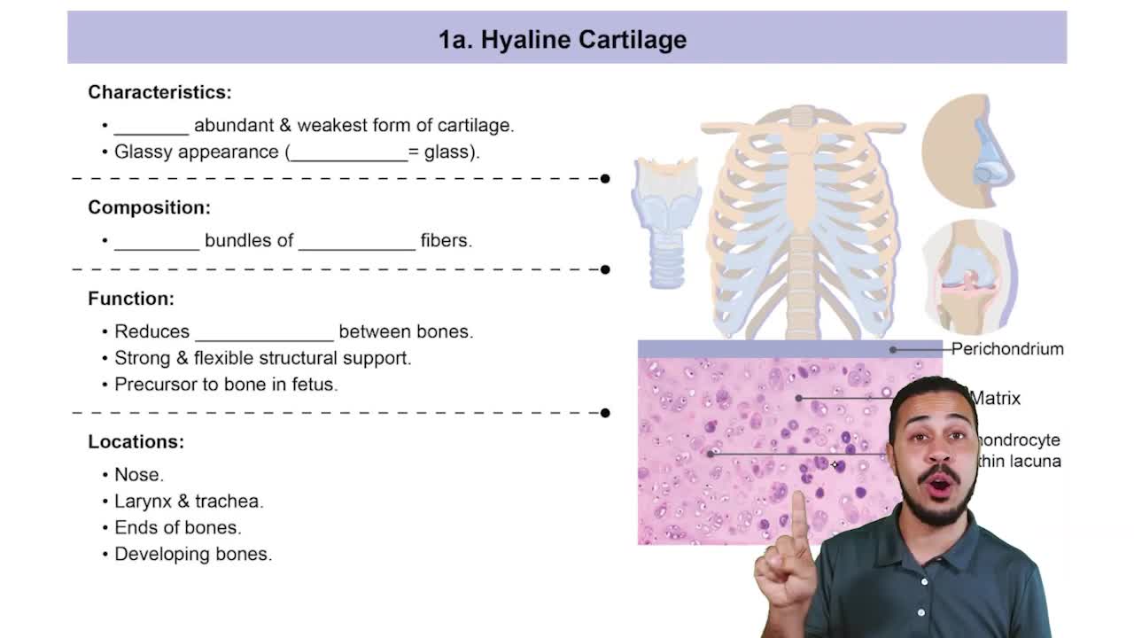
Hyaline
(most abundant),
elastic
(flexible, in ear and epiglottis), and
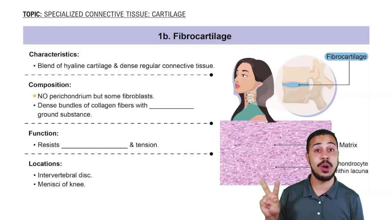
fibrocartilage
(withstands pressure, in knee and intervertebral discs).
진행 상황 추적
컨트롤 버튼이 '내비게이션' 모드로 변경되었습니다.
1/26
추천 영상
VideoThumbView.guidedCourse
01:27
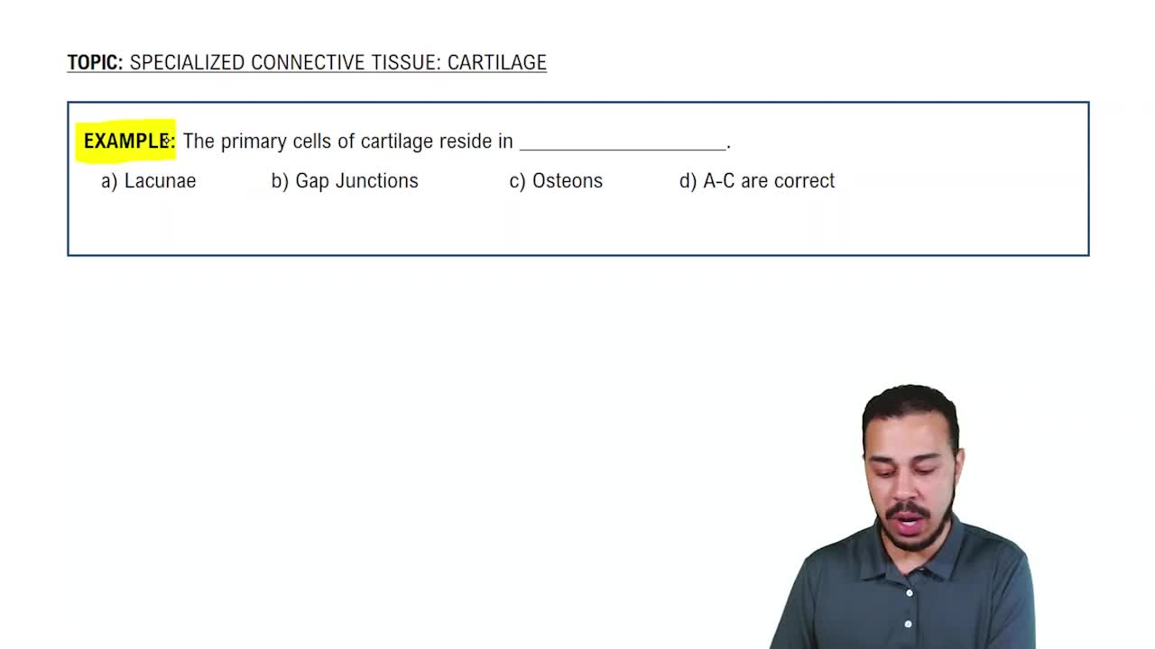
Specialized Connective Tissue: Cartilage Example 1
4893
views
66
rank
VideoThumbView.guidedCourse
05:36
Hyaline Cartilage
6397
views
100
rank
VideoThumbView.guidedCourse
05:20
Fibrocartilage
5681
views
90
rank
1
comments
이 집합의 용어 (26)
하이드의 정의
Types of skeletal cartilage
Hyaline
(most abundant),
elastic
(flexible, in ear and epiglottis), and
fibrocartilage
(withstands pressure, in knee and intervertebral discs).
Perichondrium
Dense irregular connective tissue layer that surrounds skeletal cartilage, providing support and nutrients.
Appositional cartilage growth
Growth by adding new matrix on the outer surface, causing outward expansion.
Interstitial cartilage growth
Growth from within the cartilage by division of chondrocytes inside lacunae and secretion of new matrix.
Seven functions of bones
Support, protection, anchorage, mineral storage, blood cell formation, triglyceride storage, and hormone production.
Axial vs appendicular skeleton
Axial: skull, vertebral column, rib cage (support/protection). Appendicular: limbs and girdles (movement and manipulation).
Bone shape classifications
Long (limbs), short (carpals, tarsals), flat (skull, ribs), irregular (vertebrae, hip bones).
Compact bone
Dense outer layer of bone that appears smooth and solid.
Spongy bone
Internal bone with trabeculae (needle-like pieces) that provide strength and house marrow.
Structure of a long bone
Diaphysis (shaft with medullary cavity), epiphyses (ends with spongy bone), periosteum outside, endosteum inside.
Epiphyseal line
Remnant of the epiphyseal plate (hyaline cartilage) that allows lengthwise bone growth.
Red bone marrow location
In trabecular cavities of spongy bone and diploë of flat bones; in adults, mainly in proximal epiphyses of humerus and femur.
Five types of bone cells
Osteoprogenitor, osteoblasts, osteocytes, bone lining cells, and osteoclasts.
Osteon (Haversian system)
Structural unit of compact bone, consisting of concentric lamellae around a central canal.
Canaliculi function
Tiny channels connecting lacunae and central canal, allowing osteocyte communication and nutrient exchange.
Organic vs inorganic bone components
Organic: cells and osteoid (collagen, ground substance) for flexibility. Inorganic: hydroxyapatites (calcium phosphate) for hardness.
Endochondral ossification
Bone replaces hyaline cartilage; forms most bones below the skull except clavicles.
Intramembranous ossification
Bone forms directly from fibrous connective tissue membranes; forms cranial bones and clavicles.
Zones of epiphyseal plate during bone length growth
Resting, proliferation, hypertrophic, calcification, and ossification zones.
Hormonal regulation of bone growth
Growth hormone and thyroid hormone stimulate growth; testosterone and estrogen cause growth spurts and plate closure.
Bone remodeling process
Balance of bone resorption by osteoclasts and bone deposition by osteoblasts to maintain strength and calcium homeostasis.
Role of parathyroid hormone (PTH) in bone remodeling
Stimulates osteoclasts to resorb bone, releasing calcium into the blood.
Wolff's law
Bone grows or remodels in response to the mechanical stresses it experiences.
Four stages of fracture repair
Hematoma formation, fibrocartilaginous callus formation, bony callus formation, and bone remodeling.
Osteomalacia and rickets
Disorders of inadequate bone mineralization; rickets occurs in children, osteomalacia in adults.
Osteoporosis
Condition where bone resorption exceeds formation, leading to porous, fragile bones; common in postmenopausal women.
 뒤로뒤로
 01:27