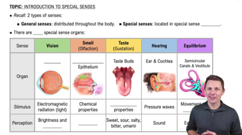
Olfactory receptors are bipolar neurons located in the nasal epithelium, specifically the pseudostratified ciliated columnar epithelium in the superior nasal cavity.
Requirements for substances to be smelled
Substances must be volatile, water-soluble, and lipid-soluble to be smelled; odorant gases must dissolve in mucus.
Neural pathway of olfactory impulses
Olfactory nerve axons synapse with mitral cells in olfactory bulbs; mitral cell axons form olfactory tracts that project to the temporal lobes and limbic system.
Function of olfactory (Bowman’s) glands
They produce mucus located in connective tissue deep to the nasal epithelium to trap and dissolve odorants.
Location of gustatory receptors
Gustatory receptor cells are located in taste buds on the tongue and other areas.
Primary tastes
The four primary tastes are sour, salty, bitter, and sweet.
Neural pathways for taste sensation
Taste impulses travel via cranial nerves VII (anterior 2/3 tongue), IX (posterior 1/3 tongue), and X (epiglottis and pharynx) to the medulla, thalamus, and parietal lobe.
Accessory structures of the eye
Include eyebrows, eyelids, eyelashes, lacrimal apparatus, and extrinsic eye muscles.
Function of the conjunctiva
A thin mucous membrane lining the inner eyelids and anterior eyeball surface, protecting and lubricating the eye.
Components of the fibrous tunic of the eye
Includes the posterior sclera, which provides shape and protection, and the anterior cornea, which is transparent and refracts light.
Role of the scleral venous sinus (canal of Schlemm)
Drains excess aqueous humor from the anterior chamber to maintain intraocular pressure.
Functions of the vascular tunic
Includes the choroid (absorbs stray light and nourishes retina), ciliary body (produces aqueous humor and controls lens shape), and iris (regulates light entry).
Structure and function of the iris
Colored part of the eye with circular and radial muscles controlling pupil size to regulate light entering the eye.
Layers of the retina
Consists of pigmented epithelium (absorbs stray light) and neural portion with photoreceptors (rods and cones), bipolar neurons, and ganglion neurons.
Differences between rods and cones
Rods detect black-and-white vision in dim light; cones detect color and sharpness in bright light, concentrated in the fovea centralis.
Function of the lens
A nonvascular, transparent structure that fine-tunes focusing of light rays for clear vision.
Cause and effect of glaucoma
Blockage of aqueous humor drainage increases intraocular pressure, damaging the retina and causing blindness.
Accommodation in vision
Ciliary muscle changes lens curvature to focus on near (thicker lens) or far (flatter lens) objects.
Process of image formation on the retina
Light rays are refracted by cornea and lens, focused inverted and reversed on retina, then corrected by the brain for perception.
Photopigments in rods and cones
Rods contain rhodopsin; cones contain three types of iodopsins for red, green, and blue color detection.
Pathway of visual impulses
From photoreceptors to bipolar cells, then ganglion cells; impulses travel via optic nerve, chiasma, tract, thalamus, to occipital cortex.
Anatomical divisions of the ear
External ear (auricle, canal, tympanic membrane), middle ear (tympanic cavity with ossicles), and internal ear (bony and membranous labyrinth).
Function of the ossicles
Malleus, incus, and stapes transmit vibrations from tympanic membrane to the oval window of the cochlea.
Role of the cochlea
Contains the organ of Corti, the receptor organ for hearing, converting sound waves into nerve impulses.
Function of the semicircular canals
Contain receptors for dynamic equilibrium, detecting rotational movements of the head.
Static equilibrium organs
Maculae of the utricle and saccule detect head position relative to gravity using otolithic membrane weighted by otoliths.
Dynamic equilibrium mechanism
Movement of endolymph in semicircular ducts bends crista ampullaris hair cells, triggering receptor potentials for balance.
Auditory pathway to the brain
Impulses from cochlear nerve pass to medulla, cross sides, then to midbrain, thalamus, and auditory cortex in temporal lobe.
 뒤로
 5:05