 뒤로
Anatomy & Physiology Test 3 Review
 04:36

이 집합의 용어 (29)
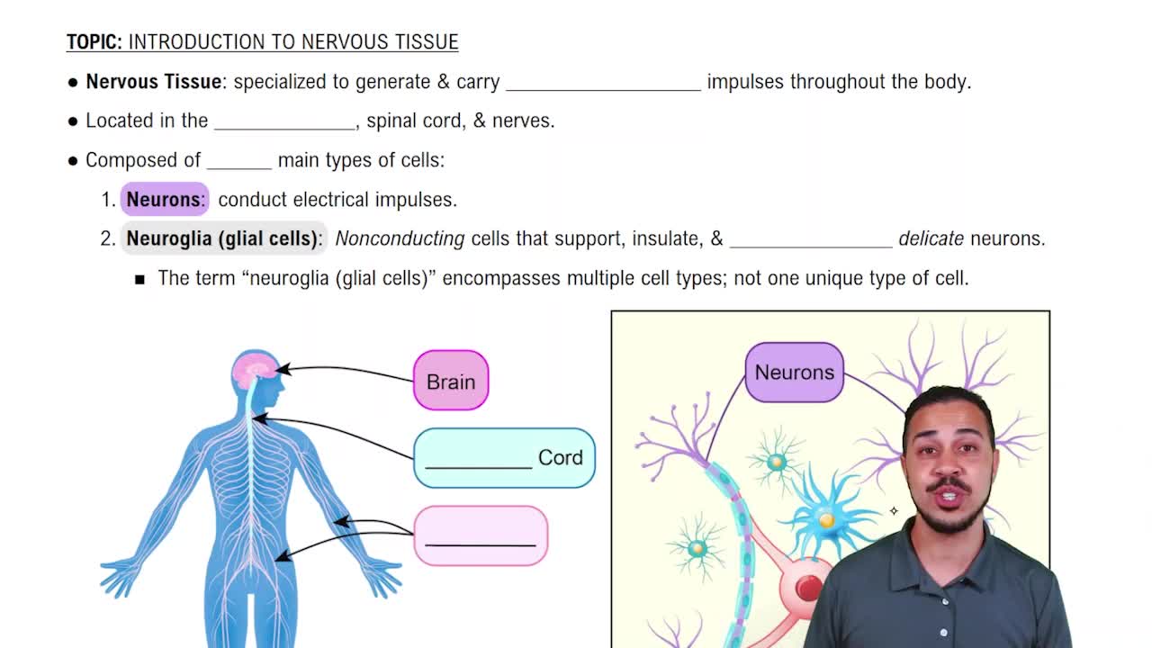
Support cells in the nervous system that maintain homeostasis, form myelin, and provide support and protection for neurons.
Membrane-bound sacs located in the axon terminals that store and release neurotransmitters into the synaptic cleft.
Endoneurium surrounds individual axons, perineurium surrounds fascicles, and epineurium surrounds the entire nerve.
In the CNS, myelin is formed by oligodendrocytes; in the PNS, by Schwann cells. CNS myelin sheaths can myelinate multiple axons, PNS sheaths one axon each.
Cranial nerves are classified as sensory, motor, or mixed. They originate from the cerebrum, midbrain, pons, or medulla oblongata.
White matter contains myelinated axons; gray matter contains neuron cell bodies and dendrites. Their distribution differs in CNS and PNS.
A selective barrier formed by endothelial cells and astrocytes that protects the brain by restricting passage of substances from blood to brain tissue.
Presynaptic neurons originate in the CNS and synapse in autonomic ganglia; postsynaptic neurons extend from ganglia to target organs.
Includes the thalamus, hypothalamus, and epithalamus, which regulate sensory relay, autonomic functions, and endocrine activity.
The outer layer of the cerebrum composed of gray matter responsible for higher brain functions like sensation, voluntary movement, and cognition.
Clusters of gray matter deep in the cerebrum involved in regulating voluntary motor control and procedural learning.
Includes the midbrain, pons, and medulla oblongata, which control vital functions and serve as pathways for nerve tracts.
Networks of intersecting nerves: cervical, brachial, lumbar, and sacral plexuses that innervate limbs and trunk.
Sensory tracts carry impulses to the brain; motor tracts carry impulses from the brain. Naming follows origin, destination, and function.
Includes receptor, sensory neuron, integration center, motor neuron, and effector.
Vitreous humor is a gel filling the posterior chamber; aqueous humor is a watery fluid in the anterior chamber.
Cataracts are clouding of the lens; glaucoma is increased intraocular pressure damaging the optic nerve.
The lens is held by suspensory ligaments connected to the ciliary body, which controls lens shape for focusing.
Rods detect light intensity and are sensitive in low light; cones detect color and function in bright light.
A major nerve from the cervical plexus that controls the diaphragm for breathing.
Somatic neurons control voluntary muscles with a single motor neuron; autonomic neurons control involuntary organs with two neurons (pre- and postganglionic).
The sympathetic division prepares for 'fight or flight'; the parasympathetic division promotes 'rest and digest'.
ANS neurons innervate organs to regulate functions like heart rate, digestion, and respiratory rate.
Four interconnected cavities filled with cerebrospinal fluid: two lateral ventricles, third ventricle, and fourth ventricle.
Includes the outer ear, middle ear with ossicles, and inner ear with cochlea and semicircular canals for hearing and balance.
The three meninges are dura mater, arachnoid mater, and pia mater. The falx cerebri is a dural fold separating cerebral hemispheres.
Gray matter in the spinal cord is shaped like a butterfly with dorsal (sensory) and ventral (motor) horns.
Includes the conus medullaris (tip of cord), filum terminale (fibrous extension), and cauda equina (nerve roots).
Cerebrospinal fluid flows from lateral ventricles to third, then fourth ventricle, into subarachnoid space, and is absorbed into venous blood.