 뒤로
Anatomy & Physiology: The Skeletal System
 3:56

이 집합의 용어 (40)
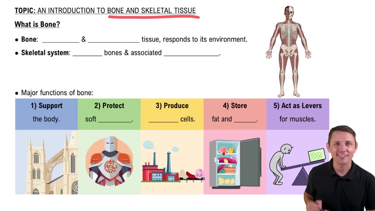
Support, protection, movement, mineral storage, blood cell formation, and energy storage.
Hyaline cartilage (nose, trachea), elastic cartilage (ear), and fibrocartilage (intervertebral discs).
Appositional growth: growth from the outside; interstitial growth: growth from within the cartilage.
Axial skeleton: skull, vertebral column, rib cage; appendicular skeleton: limbs and girdles.
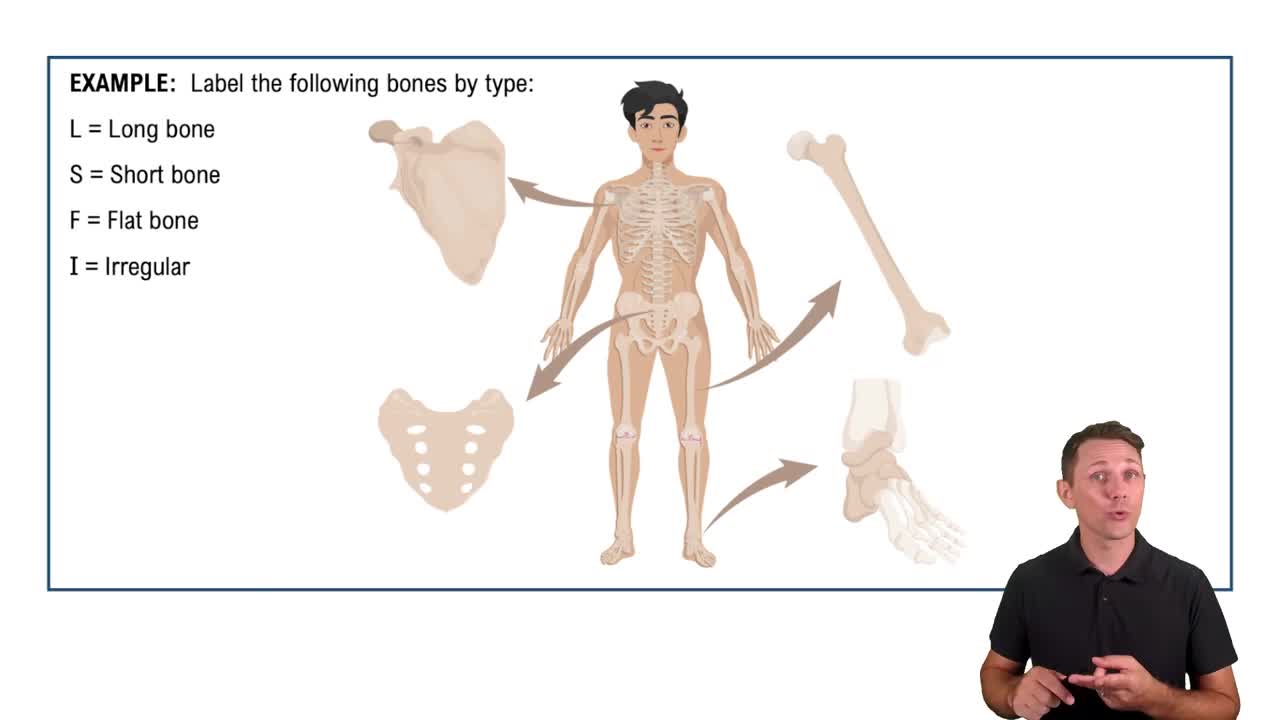
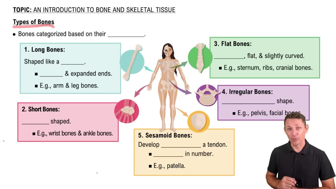
Long, short, flat, and irregular bones based on shape.
Include projections (sites of muscle/ligament attachment), surfaces (joint formations), and depressions/openings (passage for vessels/nerves).
Compact bone: dense outer layer; spongy bone: porous inner network.
Diaphysis: shaft of long bone, mainly compact bone; epiphysis: ends, mainly spongy bone with a thin compact bone layer.
Periosteum: outer fibrous layer; endosteum: inner lining of medullary cavity.
Consist of spongy bone sandwiched between two thin layers of compact bone, unlike long bones with a diaphysis and epiphyses.
Osteogenic cells (stem cells), osteoblasts (bone-forming), osteocytes (mature bone cells), osteoclasts (bone-resorbing).
The basic structural unit of compact bone, consisting of concentric lamellae surrounding a central canal.
Lamellae are concentric rings of bone matrix; types include concentric, interstitial, and circumferential lamellae.
Runs longitudinally through osteons, containing blood vessels and nerves.
Channels that run perpendicular to osteons, connecting blood vessels and nerves between central canals.
Central canals run longitudinally inside osteons; Volkmann’s canals run transversely connecting osteons.
Small cavities within bone matrix that house osteocytes.
Microscopic canals connecting lacunae, allowing nutrient and waste exchange between osteocytes.
These microscopic features are characteristic of compact bone.
Trabeculae are the lattice-like network of bone found in spongy bone.
Osteoid (collagen fibers and ground substance) produced by osteoblasts, providing tensile strength and flexibility.
Mineral salts, mainly hydroxyapatite (calcium phosphate crystals), providing hardness and resistance to compression.
Ossification is the process of bone tissue formation.
Intramembranous ossification (bone forms directly from mesenchyme) and endochondral ossification (bone replaces hyaline cartilage).
Mesenchymal cells differentiate into osteoblasts, secrete osteoid, which calcifies, forming woven bone and then lamellar bone.
Hyaline cartilage model forms, cartilage calcifies, primary ossification center develops in diaphysis, secondary centers in epiphyses, cartilage replaced by bone.
Interstitial growth (lengthwise at epiphyseal plates) and appositional growth (widthwise by adding bone tissue).
Resting zone, proliferation zone, hypertrophic zone, calcification zone, and ossification zone.
Growth hormone, thyroid hormone, sex hormones (estrogen and testosterone), and calcitonin.
Deposition: osteoblasts build bone; resorption: osteoclasts break down bone.
Maintain bone strength, repair damage, and regulate blood calcium levels.
Osteoblasts synthesize and secrete bone matrix.
Osteoclasts break down bone matrix by secreting acids and enzymes.
Essential for bone strength, muscle contraction, nerve transmission, and blood clotting.
Parathyroid hormone (PTH) increases blood calcium by stimulating osteoclasts; calcitonin lowers blood calcium by inhibiting osteoclasts.
Bone remodels in response to mechanical stress, becoming stronger where stressed.
Simple, compound, comminuted, greenstick, spiral, and compression fractures.
Hematoma formation, fibrocartilaginous callus formation, bony callus formation, and bone remodeling.
Include osteoporosis, osteomalacia, Paget’s disease, and rickets.
Age, gender (female), low calcium/vitamin D, sedentary lifestyle, smoking, and hormonal changes.