 뒤로
Anatomy & Physiology(CH. 6-A): Skeletal System and Bone Structure
 3:56

이 집합의 용어 (39)
layer of dense connective tissue surrounding cartilage that helps resist outward expansion and contains blood vessels for nutrient delivery.
-crtilage is made up of chondrocytes (cells encased in small cavaties-lacunae)
Name the 2/3 types of cartilage and a key feature of each. (elastic & fibro)
Elastic cartilage: contains elastic fibers, found in external ear and epiglottis; Fibrocartilage: thick collagen fibers, great tensile strength, found in menisci and vertebral discs.
appositional growth (new matrix laid on surface) and interstitial growth (chondrocytes w/in lacunea divide and secrete matrix from within-new matrix within cartilage)
-casification of cartilage occurs during normal bone growth in youth, can in old age too
-hardened cartilage not the same as bone
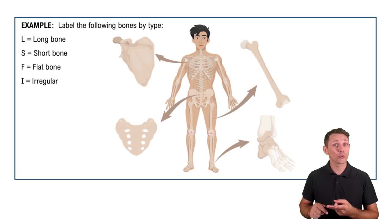
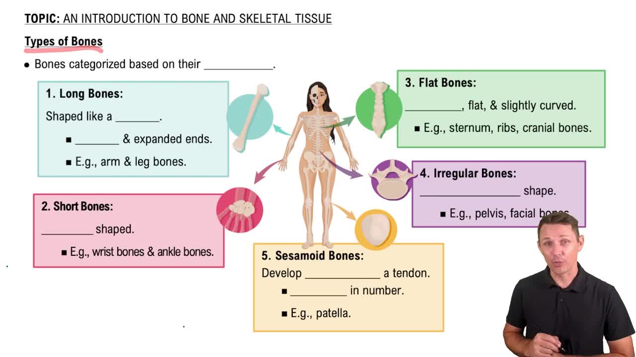
Long bones: longer than wide, e.g., limbs; Short bones: cube-shaped, e.g. wrist bones (contain sesamoid bones: form w/in tendons-patella--vary in type in people; Flat bones: thin/flat and curved, e.g. scapula; Irregular bones: complicated shapes, e.g. vertebrae.
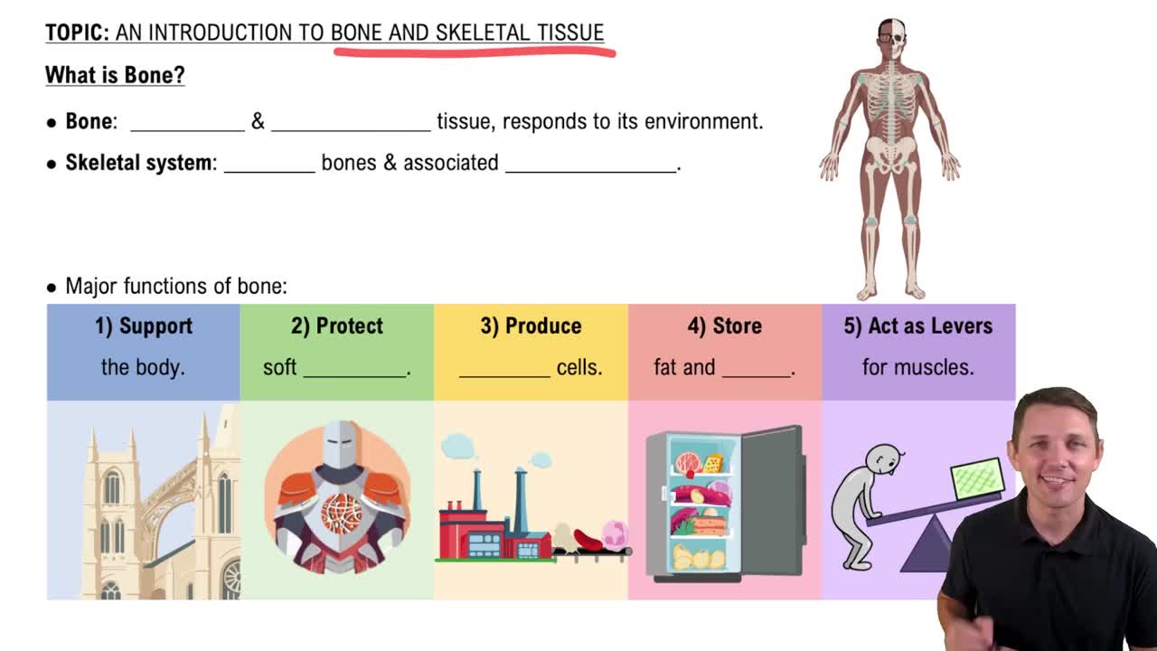
What are the seven functions of bones? (Support, protection, movement, mineral and growth factor storage (calcium/phosphorus & growth factors reservoir)
blood cell formation (hematopoiesis occurs in red bone marrow), triglyceride storage (fat stored in bone cavities), and hormone production (osteocalcin secreted by bones helps to regulate insulin secretion, glucose levels, and metabolism)
Bones contain multiple tissue types including osseous tissue, nervous tissue, cartilage, fibrous connective tissue, muscle cells, and epithelial cells.
-3 levels of structure: Gross, Microscopic, Chemical
Compact bone is dense and smooth forming the outer layer; spongy bone consists of a honeycomb, needle-like or flat trabeculae with spaces filled with red/yellow bone marrow.
A long bone has a shaft (diaphysis), bone ends (epiphyses), & membranes.
-Dia: tubular shaft that forms lost axis of bone-consists of vompact bone surrounding central medullary cavity filled with yellow marrow in adults.
Periosteum: white double-layered membrane covering external bone surfaces except joints; endosteum: delicate connective tissue membrane covering internal bone surfaces and trabeculae.
Explain Hemapoetic tissue in bones
Red marrow is found in trabecular cavities of spongy bone and flat bone diploë; it is the site of hematopoiesis (blood cell formation).
-yellow can convert into red, if become anemic
-newborns: medullary cavities and all spongey bone contain red marrow. adults: in femur/humerus and what's said above.
Bone markings are sites of muscle, ligament, and tendon attachment or joint formation/conduits for blood vessels & nerves. Types: projections: outward bulge of bone, depressions: bowl/groove-like cut-out, and openings: hole/canal (De & Op: passageways for blood cells and nerves).
specialized form of the same basic cell type. Osteogenic cells, osteoblasts, osteocytes, bone-lining cells, and osteoclasts.
Osteoblasts are bone-forming cells that secrete unmineralized bone matrix called osteoid, which contains collagen and calcium-binding proteins (collagen makes up 90% of bone protein). actively mitotic.
Osteocytes are mature bone cells in lacunae that no longer divide that maintain bone matrix, act as stress/strain sensors (such as increased force on bone or weightlessness). and communicate with osteoblasts and osteoclasts for remodeling.
Derived from same hematopoietic stem cells that become macrophages.they are large multinucleate cells that resorb (break down) bone by secreting enzymes, aiding in bone remodeling. When active: located in depressions called resorption bays. have ruffled borders.
An osteon is the structural unit of compact bone. consists of an elongated cylinder that runs parallel to long axis of bone (acts as tiny-weight-bearing pillars.
Hair-like canals connecting lacunae to eachother and central canal. When matrix hardens & cells are trapped the canaliculi form: allowing osteocytes to communicate and exchange nutrients and wastes.
What are the organic compounds of bone?
Organic osteogenic cells, osteoblasts, osteocytes, bone-lining cells, osteoclasts, and osteoid (makes up 1/3 of organic bone matrix, is secreted by osteoblasts). ground substance & collagen fibers -high tensil strength & flexibility of bone.
Explain Hyaline Cartilage
-most abundant type, offers flexible support/resilience
-contains collagen fibers only
-articular (joints), costal (ribs), respiratory (larynx), nasal cartilage (nose tip)
Explain the structure of short, irregular, & flat bones
-thin plates of spongey bone (dipole) covered by compact bone. compact bone sandwitched b/t connective tissue memrbanes.
-Bone marrow scattered throughout spongy bone, no defined marrow cavity.
-flat bones: layer of spongy bone sandwiched b/t 2 thin layers of compact bone
Explian Periosteum & Endosteum & Hyaline cartilage
P: covers outside of compact bone, E: covers inside portion of compact bone
-H: covers area of bone that is part of a movable joint
Explain Epiphyses & and Epiphyses line of longs bones
Epi: ends of long bones, consist of compact bone externally & spngey bone internally
-articular cartilage covers articular (joint) surfaces
B/t disphysis & epiphyses: epiphyseal line =epiphyseal plate where bone growth occurs
Explain more in depth the periosteum
Fibrous layer: outer layer consisting of dense irregular connective tissue consisting of Sharpey's fibers that secure bone matrix.
Osteogenic layer: inner layer abutting bone & contains primitive osteogenic stem cells that gives rise to most all bone cells
Nutrient Foramen of Periosteum?
the periosteum contains many nerve fibers/ blood vessels that continue on to the shaft through these openings.
-the periosteum has anchoring points for tendons and ligaments
Explian Endosteum more in depth
-covers trabeculae of spongy bone
-lines canals that pass through compact bone
-like periosteum, contains osteogenic cells that can differentiate into other bone cells
Eplain Osteogenic cells
also called osteoprogenitor cells. Mitotically active stem cells in periosteum & active endosteum. When stimulated, they turn into osteoblasts or bone-lining cells . some remain as osteogenic stem cells
Explain Bone-lining cells
help maintain matrix. On external bone surface: periosteal cells . On internal surfaces: endosteal cells
Explain compact bone
also called lamellar bone.
consists of osteon (Haversian system), canals & canaliculi, and interstitial & circumferential lamallae
Explain Lamallae
an osteon cylinder consists of several rings of bone matrix called this.
-contain collagen fibers that run in different directions in adjacent rings
-withstands stress & resist twisting
-bone salts are found b/t collagen fibers
Explain Central (Haversian) canal & Perforating (Volkmann's) canals
Cen: runs through core of osteon. contains blood vessels & nerve fibers. Per: canals lined w/ endosteum that occur at right angles to central canal. connects blood vessels & nerves of periosteum, medullary cavity, & central canal.
What are Lacunae ?
small cavities that contain osteocytes
-osteoblasts that secrete bone matrix maintain contact w/ each other & osteocytes via cell projections w/ gap junctions
Explain Interstitial Lamallae
lamaellae that aren't part of osteon
-some fill gaps b/t forming osteons; others are remnants of osteons cut by bone remodeling
Explain Circumferential lamallae
-just deep to periosteum, but superficial to endosteum, these layers of lamallae extend around entire surface of diaphysis
-help long bone to resist twisting
Explain spongy bone
appears poorly organized, but is along lines of stress to help bone resist stress
-trabeculae, like cables on a suspension bridge, confer strength to bone
no osteons. but trab. have irregularly arranged lamalae & osteocytes interconnected by canaliculi. capillaries in endos. supply nutrients
Explain Sacrificial bonds
resisience of bone is due to this. in or b/t collagen molecules that stretch & break to dissipate energy & prevent fracutures. if no additional trauma, bonds re-form
What are the inorganic componets of bone?
Hydroxyapatites (mineral salts)-65% of bone mass. mainly tiny Ca phosphate crystals in & around collagen fibers. hardness & resistance to compression
Why is bone important?
half as strong as steel when resisting compression. just as strong when resisting tension
-lasts long b/c of mineral composition
-forensic scientists can look at bones and see evidence of trauma which can be caried in bones for decades