Skip to main content
Anatomy & Physiology
나의 코스
배우기
시험 준비
AI 튜터
학습 가이드
교과서적 해결책
플래시카드
탐험해 보세요
앱을 사용해 보세요
나의 코스
배우기
시험 준비
AI 튜터
학습 가이드
교과서적 해결책
플래시카드
탐험해 보세요
앱을 사용해 보세요
뒤로
Axial Muscles of the Body - Anatomy & Physiology
카드를 뒤집기 위해 탭할 수 있습니다.
Muscles of facial expression
카드를 뒤집기 위해 탭할 수 있습니다.
👆
Muscles of facial expression
Thin muscles in the face and scalp that often insert into skin, innervated by the facial nerve (cranial nerve VII).
진행 상황 추적
컨트롤 버튼이 '내비게이션' 모드로 변경되었습니다.
1/28
추천 영상
VideoThumbView.guidedCourse
6:31
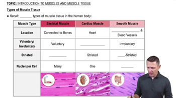
Types of Muscle Tissue
18732
views
494
rank
1
comments
VideoThumbView.guidedCourse
2:00
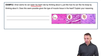
Introduction to Muscles and Muscle Tissue Example 1
16657
views
324
rank
VideoThumbView.guidedCourse
4:36
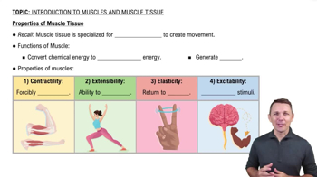
Properties of Muscle Tissue
25638
views
741
rank
2
comments
이 집합의 용어 (28)
하이드의 정의
Muscles of facial expression
Thin muscles in the face and scalp that often insert into skin, innervated by the facial nerve (cranial nerve VII).
Epicranius muscle
Has frontal belly that raises eyebrows and wrinkles forehead, and occipital belly at the back of the head.
Orbicularis oculi action
Closes the eyelids during blinking.
Zygomaticus major function
Extends from the zygomatic region to the mouth and produces a smile.
Buccinator muscle role
Forms the muscular wall of the cheek and compresses the cheek during chewing.
Innervation of muscles of mastication
Mandibular division of the trigeminal nerve (cranial nerve V).
Masseter muscle function
Prime mover of jaw closure, located over the lateral ramus of the mandible.
Extrinsic tongue muscles innervation
Innervated by the hypoglossal nerve (cranial nerve XII) and move the tongue in various directions.
Suprahyoid muscles
Located superior to the hyoid bone; include mylohyoid, stylohyoid, and digastric muscles.
Infrahyoid muscles
Located inferior to or associated with the hyoid and laryngeal cartilages; include sternohyoid, omohyoid, sternothyroid, and thyrohyoid.
Sternocleidomastoid muscle action
Flexes the head and neck and divides the neck into anterior and posterior triangles.
Erector spinae muscle group
Largest deep back muscle group with three columns: iliocostalis, longissimus, and spinalis, responsible for trunk extension.
Diaphragm function
Primary muscle of respiration; contraction flattens it to increase thoracic cavity volume.
External intercostals action
Lift the rib cage during inspiration (breathing in).
Internal intercostals action
Assist expiration during heavy breathing by depressing the rib cage.
Layers of the abdominal wall muscles
From superficial to deep: external oblique, internal oblique, transversus abdominis, and rectus abdominis.
Linea alba
Midline fibrous seam formed by interlacing abdominal aponeuroses; insertion site for rectus abdominis.
Pelvic diaphragm composition
Consists of levator ani (iliococcygeus, puborectalis, pubococcygeus) and coccygeus muscles; supports pelvic organs.
Urogenital diaphragm muscles
Includes sphincter urethrae (external urethral sphincter) and deep transverse perineus muscles.
Superficial perineal muscles
Include ischiocavernosus, bulbospongiosus, and superficial transverse perineus muscles.
Pectoralis major muscle
Large superficial muscle covering the upper anterior thorax involved in scapular movement.
Pectoralis minor muscle
Smaller, deep muscle beneath pectoralis major, exposed when pectoralis major is removed.
Serratus anterior muscle
Saw-toothed muscle slips along the lateral ribs, involved in scapular movement.
Distinction between facial expression and mastication muscles
Facial expression muscles insert into skin and use CN VII; mastication muscles use mandibular division of CN V.
Difference between buccinator and masseter
Buccinator compresses the cheek; masseter closes the jaw.
Omohyoid muscle bellies
Has superior and inferior bellies, part of the infrahyoid muscle group.
Digastric muscle bellies
Has anterior and posterior bellies, part of the suprahyoid muscle group.
Central tendon of the diaphragm
Nonmuscular central region where diaphragm muscle fibers converge.
 뒤로뒤로
 6:31