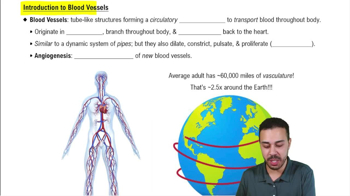
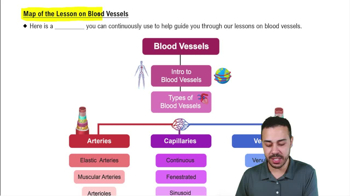
Arteries carry blood away from the heart, veins carry blood back to the heart, and capillaries connect smallest arteries to smallest veins to create a circuit.
Layers of blood vessel walls
Blood vessel walls have three tunics: tunica interna (endothelium lining), tunica media (smooth muscle and elastic tissue), and tunica externa (loose connective tissue).
Function of tunica interna
Lines the vessel, acts as a selectively permeable barrier, secretes chemicals for dilation/constriction, and normally repels blood cells to prevent clotting.
Function of tunica media
Consists of smooth muscle and elastic tissue; controls vessel diameter by contraction and strengthens vessels to prevent rupture.
Function of tunica externa
Outermost layer of loose connective tissue; anchors vessels and provides passage for nerves and lymphatic vessels; contains vasa vasorum in large vessels.
Classes of arteries by size
Conducting (large) arteries like the aorta, distributing (medium) arteries to organs, and resistance (small) arteries including arterioles that control blood flow.
Aneurysm
A weak point in an artery or heart wall forming a bulging sac that may rupture; caused by atherosclerosis, hypertension, trauma, or infection.
Arterial sense organs
Sensory structures in vessel walls that monitor blood pressure and chemistry, including carotid sinuses (baroreceptors) and carotid/aortic bodies (chemoreceptors).
Types of capillaries
Continuous (most tissues), fenestrated (rapid absorption organs), and sinusoids (liver, bone marrow) distinguished by permeability.
Capillary beds and precapillary sphincters
Networks of capillaries controlled by sphincters that regulate blood flow; sphincters relax to perfuse capillaries and contract to bypass them.
Veins as capacitance vessels
Veins have thin walls, collapse when empty, expand easily, and contain about 64% of blood at rest; they have valves to prevent backflow.
Types of veins
Postcapillary venules, muscular venules, medium veins (with valves), large veins, and venous sinuses (thin walls, no smooth muscle).
Varicose veins
Enlarged, twisted superficial veins caused by valve failure, blood pooling, and vessel wall weakening; risk factors include heredity, obesity, and pregnancy.
Simplest circulatory route
Heart → arteries → arterioles → capillaries → venules → veins; blood passes through one capillary network per circuit.
Blood flow and perfusion
Flow is blood volume per time (mL/min); perfusion is flow per tissue mass (mL/100g/min); cardiac output at rest is about 5.25 L/min.
Blood pressure (BP) measurement
Measured at brachial artery; systolic pressure is peak during ventricular contraction; diastolic pressure is minimum during relaxation; normal ~120/75 mm Hg.
Resistance depends on blood viscosity, vessel length, and vessel radius; radius has greatest effect, with flow proportional to radius to the fourth power.
Vasomotion
Collective term for vasoconstriction and vasodilation; regulates blood pressure and flow by changing vessel radius.
Regulation of blood pressure and flow
Controlled by local (autoregulation), neural (vasomotor center reflexes), and hormonal mechanisms (angiotensin II, aldosterone, ADH, catecholamines).
Capillary exchange mechanisms
Substances cross capillary walls by diffusion, transcytosis, filtration, and reabsorption through endothelial cells, intercellular clefts, and fenestrations.
Filtration and reabsorption in capillaries
Fluid filters out at arterial end due to hydrostatic pressure and is reabsorbed at venous end due to colloid osmotic pressure; about 85% reabsorbed.
Edema causes
Excess fluid accumulation due to increased filtration, reduced reabsorption, or lymphatic obstruction; can impair oxygen delivery and cause tissue damage.
Mechanisms of venous return
Venous return aided by pressure gradient, gravity, skeletal muscle pump, thoracic pump, and cardiac suction.
Skeletal muscle pump
Contraction of limb muscles compresses veins, pushing blood toward the heart; valves prevent backflow.
Thoracic (respiratory) pump
Inhalation lowers thoracic pressure and raises abdominal pressure, forcing blood upward in the inferior vena cava.
Effects of exercise on venous return
Exercise increases heart rate, dilates vessels, and enhances skeletal muscle and thoracic pumps, improving venous return.
Arterial pressure points
Superficial arteries close to body surface used for pulse taking and emergency pressure to reduce bleeding, e.g., femoral artery in femoral triangle.
Hypertension
Chronic resting BP >130/80 mm Hg; damages heart and vessels, promotes atherosclerosis; primary (90%) and secondary forms exist.
 뒤로
 3:38