Skip to main content
Anatomy & Physiology
나의 코스
배우기
시험 준비
AI 튜터
학습 가이드
교과서적 해결책
플래시카드
탐험해 보세요
앱을 사용해 보세요
나의 코스
배우기
시험 준비
AI 튜터
학습 가이드
교과서적 해결책
플래시카드
탐험해 보세요
앱을 사용해 보세요
뒤로
Cardiovascular System and Blood - Anatomy & Physiology
카드를 뒤집기 위해 탭할 수 있습니다.
Major functions of the cardiovascular system components
카드를 뒤집기 위해 탭할 수 있습니다.
👆
Major functions of the cardiovascular system components
Blood
transports gases, nutrients, and wastes;
heart
pumps blood;
blood vessels
carry blood throughout the body.
진행 상황 추적
컨트롤 버튼이 '내비게이션' 모드로 변경되었습니다.
1/29
추천 영상
VideoThumbView.guidedCourse
2:36
Introduction to Blood Example 1
11277
views
332
rank
VideoThumbView.guidedCourse
7:49
Composition of Blood
11822
views
336
rank
VideoThumbView.guidedCourse
1:55
Introduction to Blood Example 2
9302
views
223
rank
이 집합의 용어 (29)
하이드의 정의
Major functions of the cardiovascular system components
Blood
transports gases, nutrients, and wastes;
heart
pumps blood;
blood vessels
carry blood throughout the body.
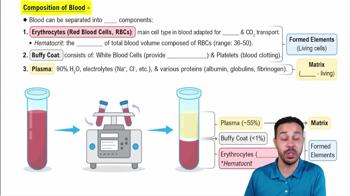
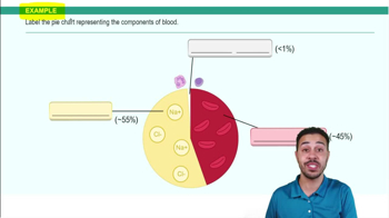
General composition of blood
Blood consists of
plasma
(liquid matrix) and
formed elements
(erythrocytes, leukocytes, platelets).
Composition of blood plasma
Plasma is mostly water with dissolved
proteins
,
electrolytes
,
nutrients
,
gases
, and
waste products
.
Major plasma proteins and their functions
Albumins
maintain osmotic pressure;
globulins
include antibodies;
fibrinogen
is essential for clotting; all produced mainly by the liver.
Formed elements of blood and their general functions
Erythrocytes
transport oxygen;
leukocytes
defend against infection;
platelets
aid in clotting.
Five types of leukocytes in order of prevalence
Neutrophils, lymphocytes, monocytes, eosinophils, basophils; each has specific immune functions.
Structure and function of hemoglobin
Hemoglobin is a protein in erythrocytes that binds oxygen; breakdown produces iron, biliverdin, and bilirubin.
Definition and normal ranges of hematocrit
Hematocrit is the percentage of erythrocytes in blood; normal ranges: males ~42-52%, females ~37-47%.
Locations of hematopoiesis
Hematopoiesis occurs mainly in red bone marrow; hematopoietic stem cells (HSCs) give rise to all blood cells.
Erythropoiesis and regulation
Erythropoiesis is red blood cell formation; reticulocytes are immature RBCs; regulated by erythropoietin (EPO) from kidneys.
Basic process of leukopoiesis
Leukopoiesis is the production of white blood cells from hematopoietic stem cells.
Basic process of thrombopoiesis
Thrombopoiesis is platelet production from megakaryocytes in bone marrow.
Vascular phase of hemostasis
Endothelial cells constrict vessels and release factors to reduce blood loss after injury.
Role of platelets in hemostasis
Platelets adhere to injury sites, aggregate, and form a platelet plug to stop bleeding.
Basic steps of coagulation
Coagulation forms fibrin clot via intrinsic, extrinsic, and common pathways activating clotting factors.
Difference between intrinsic and extrinsic coagulation pathways
Intrinsic is activated by contact with damaged vessel; extrinsic is triggered by tissue injury; both converge on common pathway.
Role of vitamin K in blood clotting
Vitamin K is essential for synthesizing clotting factors II, VII, IX, and X.
Process of fibrinolysis
Fibrinolysis dissolves clots using plasmin, activated from plasminogen by tissue plasminogen activator.
ABO blood group antigens and antibodies
Blood types have A or B antigens; antibodies are opposite type (e.g., type A has anti-B antibodies).
Rh blood group and clinical significance
Presence of Rh antigen = positive; absence = negative; anti-Rh antibodies can cause hemolytic disease in newborns.
Position of the heart in the thoracic cavity
The heart lies in the mediastinum, between the lungs, behind the sternum, and above the diaphragm.
Layers of the pericardium
Fibrous pericardium (outer), parietal serous pericardium, pericardial cavity with fluid, and visceral serous pericardium (epicardium).
Structural differences between atria and ventricles
Atria have thinner walls and receive blood; ventricles have thicker walls and pump blood out.
Primary internal heart structures
Chambers, septa, valves, papillary muscles, chordae tendineae, fibrous skeleton, and vessel openings.
Blood flow through the heart
Blood flows: body → right atrium → right ventricle → lungs → left atrium → left ventricle → body; oxygen-poor to lungs, oxygen-rich to body.
Phases of cardiac muscle action potential
Phases: rapid depolarization, plateau (Ca2+ influx), repolarization; ion movements involve Na+, Ca2+, and K+.
Electrical conduction system sequence
SA node → AV node → AV bundle → bundle branches → Purkinje fibers; coordinates heart contractions.
Definition of cardiac cycle, systole, and diastole
Cardiac cycle is one heartbeat; systole is contraction phase; diastole is relaxation phase.
Cardiac output (CO) formula
CO = stroke volume (SV) × heart rate (HR); measures blood volume pumped per minute.
 뒤로뒤로
 2:36