What are the main components of the cardiovascular system?
The cardiovascular system consists of the heart and blood vessels. The heart pumps blood, vessels deliver blood to tissues and return it to the heart.
What is the difference between arteries, veins, and capillaries?
Arteries carry blood away from the heart, veins carry blood toward the heart, and capillaries connect the smallest arteries to the smallest veins.
What are the two major divisions of the cardiovascular system?
The pulmonary circuit carries blood to the lungs for gas exchange and back to the heart. The systemic circuit supplies oxygenated blood to the body tissues and returns deoxygenated blood to the heart.
Describe the flow of blood through the pulmonary circuit.
Oxygen-poor blood arrives at the right heart, is pumped through the pulmonary trunk and arteries to the lungs, where it picks up oxygen, then returns oxygen-rich blood to the left heart via pulmonary veins.
Describe the flow of blood through the systemic circuit.
Oxygen-rich blood is pumped from the left heart through the aorta to body tissues, where oxygen is released and carbon dioxide is picked up. Deoxygenated blood returns to the right heart via the superior and inferior vena cava.
Where is the heart located and what is its general shape and size?
The heart is located in the mediastinum between the lungs. It has a wide superior base and a tapered inferior apex. It is about the size of a fist, weighing ~10 ounces in adults.
What is the pericardium and its function?
The pericardium is a double-walled sac enclosing the heart, allowing it to beat without friction, providing room to expand, and resisting excessive expansion.
Name the layers of the pericardium.
The pericardium has an outer fibrous pericardium and an inner serous pericardium, which has a parietal layer lining the fibrous layer and a visceral layer (epicardium) adhering to the heart.
What are the three layers of the heart wall?
The heart wall consists of the epicardium (outer serous membrane), myocardium (middle cardiac muscle layer), and endocardium (inner smooth lining).
What is the fibrous skeleton of the heart?
A framework of collagenous and elastic fibers that provides structural support, anchors cardiac muscle and valves, and electrically insulates atria from ventricles.
How many chambers does the heart have and what are they?
The heart has four chambers: two superior atria (right and left) that receive blood, and two inferior ventricles (right and left) that pump blood out.
What is the difference between the right and left ventricles?
The left ventricle has a wall 2-4 times thicker than the right ventricle, reflecting its greater workload pumping blood to the entire body.
What are the atrioventricular (AV) valves and their function?
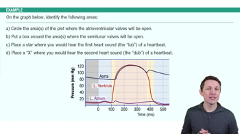
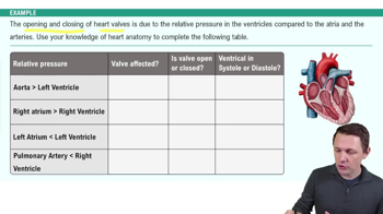
AV valves control blood flow between atria and ventricles. The right AV valve is the tricuspid valve (3 cusps), and the left AV valve is the mitral valve (2 cusps).
What are chordae tendineae and their role?
Chordae tendineae are connective tissue strings that attach AV valve cusps to papillary muscles, preventing valve prolapse during ventricular contraction.
What are the semilunar valves and their function?
Semilunar valves control blood flow from ventricles into great arteries: the pulmonary valve (right ventricle to pulmonary trunk) and the aortic valve (left ventricle to aorta).
Outline the pathway of blood flow through the heart chambers.
Blood enters right atrium → right AV valve → right ventricle → pulmonary valve → pulmonary trunk → lungs → pulmonary veins → left atrium → left AV valve → left ventricle → aortic valve → aorta → body.
What is the coronary circulation?
The heart's own blood supply system, consisting of coronary arteries branching from the aorta and coronary veins that drain into the right atrium.
What causes angina pectoris and myocardial infarction?
Angina pectoris is chest pain from partial coronary artery obstruction causing ischemia. Myocardial infarction is death of heart muscle due to prolonged obstruction.
What is the cardiac conduction system?
A system of specialized cardiomyocytes that generate and conduct electrical impulses: SA node → atria → AV node → AV bundle → bundle branches → Purkinje fibers → ventricles.
What is the function of the SA node?
The sinoatrial (SA) node is the heart's pacemaker, initiating each heartbeat and setting the heart rate by generating rhythmic electrical impulses.
What is stroke volume and how is cardiac output calculated?
Stroke volume (SV) is the amount of blood ejected per beat (~70 mL). Cardiac output (CO) = SV × heart rate (HR).
What factors affect stroke volume?
Stroke volume is influenced by preload (ventricular stretch), contractility (strength of contraction), and afterload (resistance to ejection).
How does the autonomic nervous system regulate heart rate?
The cardioacceleratory center increases HR via sympathetic nerves; the cardioinhibitory center decreases HR via parasympathetic (vagus) nerves.
What is the significance of the ECG P wave, QRS complex, and T wave?
P wave: atrial depolarization; QRS complex: ventricular depolarization; T wave: ventricular repolarization.
What causes heart sounds 'lubb' and 'dupp'?
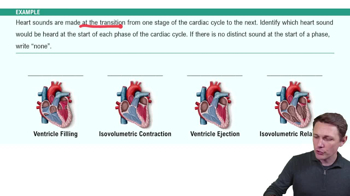
'Lubb' (S1) is caused by closure of AV valves during ventricular contraction; 'dupp' (S2) is caused by closure of semilunar valves during ventricular relaxation.
What is the Frank-Starling law of the heart?
Stroke volume is proportional to end-diastolic volume; the heart ejects as much blood as it receives due to myocardial stretch.
What is tachycardia and bradycardia?
Tachycardia is a resting heart rate above 100 bpm; bradycardia is a resting heart rate below 60 bpm.
 뒤로
 3:55