 뒤로
chapter 4
 03:32

이 집합의 용어 (25)
A microscope that transmits electrons through a thin section to view internal cell structures with resolution up to 0.2 nm.
Stratified epithelium capable of stretching and recoiling without damage, found in the urinary system.
A microscope that bounces electrons off a coated surface to create 3D images of surface features.
Exocrine glands secrete into ducts onto epithelial surfaces; endocrine glands release hormones into interstitial fluid and blood.
Structural and functional differences between the exposed apical surface and the attached basolateral surface.
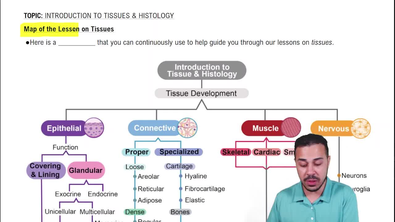
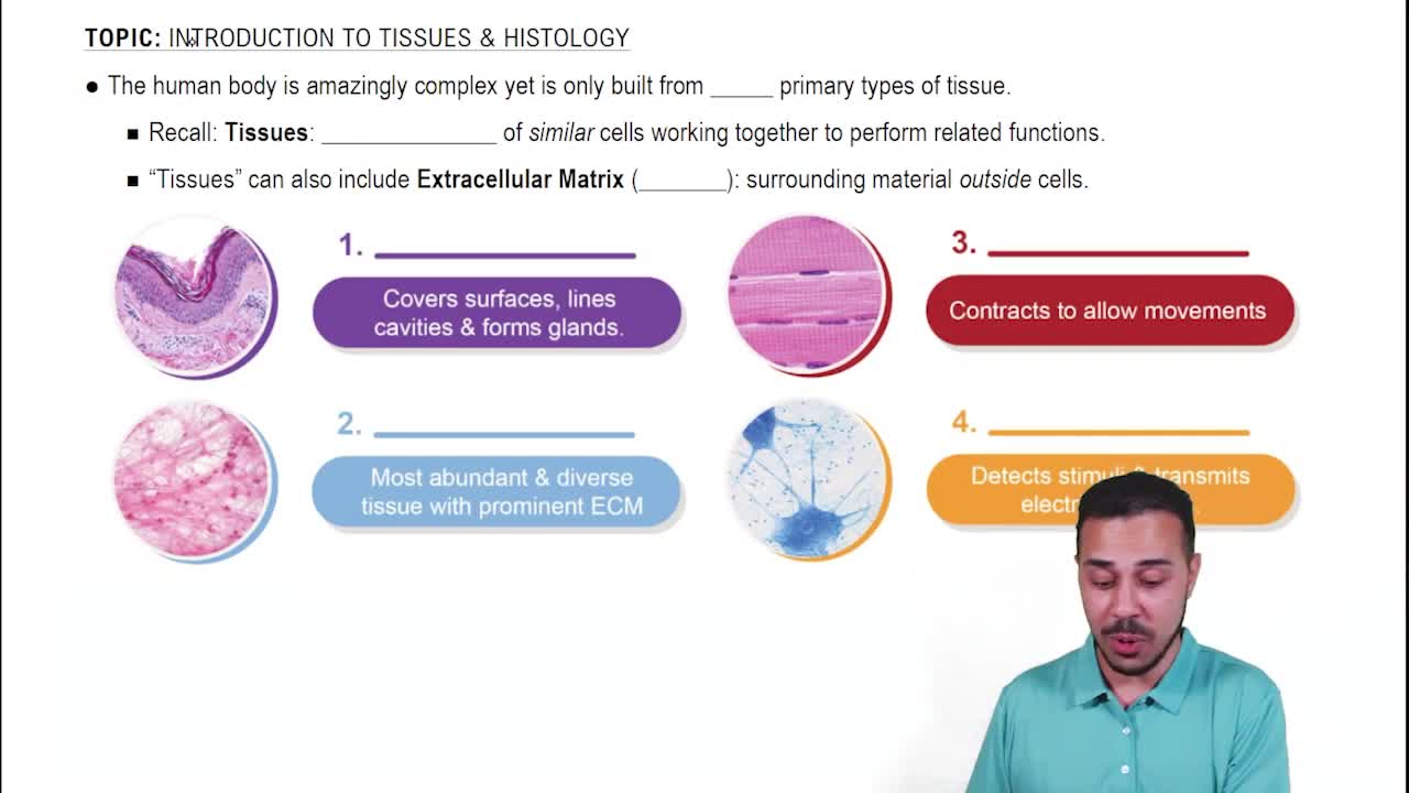
Epithelial, Connective, Muscle, and Nervous (Neural) tissues.
Increase surface area for absorption of extracellular materials, abundant in digestive and urinary tracts.
Interlocking membrane proteins that seal adjacent plasma membranes, preventing passage of water and solutes between cells.
Channels formed by connexon proteins allowing small molecules and ions to pass directly between adjacent cells.
Found in moist, high-abrasion areas like oral cavity, pharynx, esophagus, anus, and vagina.
Junctions that attach the deepest epithelial cells to the underlying basement membrane.
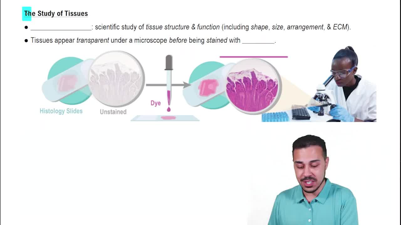
The study of tissues, focusing on microscopic groups of cells.
Secretory method where the entire cell bursts to release its product, destroying the cell (e.g., sebaceous glands).
Secretion by exocytosis without damaging the cell, common in salivary glands.
Extracellular protein fibers and clear fluid ground substance surrounding specialized cells.
Small chambers within matrix containing chondrocytes (cartilage) or osteocytes (bone).
Fixed connective tissue cells that synthesize extracellular fibers and ground substance.
Form a branching framework (stroma) supporting functional cells in organs like liver and spleen.
Single layer of columnar cells with nuclei at varying heights, often ciliated, lining trachea and nasal cavities.
Dense regular has parallel collagen fibers resisting linear tension; dense irregular has meshwork resisting multi-directional tension.
Appositional adds new matrix layers on surface; interstitial expands cartilage from within by cell division.
Membranes of mesothelium and areolar tissue lining sealed cavities, producing watery friction-reducing fluid.
Short, branched, single-nucleated striated cells joined by intercalated discs.
Supporting nervous tissue cells that maintain structure, perform phagocytosis, provide nutrients, and repair framework.
Permanent replacement of normal tissue by scar tissue when full regeneration is not possible.