뒤로

chapter 5

컨트롤 버튼이 '내비게이션' 모드로 변경되었습니다.
1/31
  • Two major components of the integumentary system

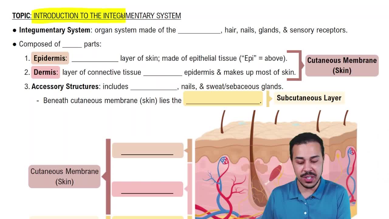
    Cutaneous membrane (Epidermis + Dermis) and Accessory structures (hairs, nails, glands, sensory receptors, nerve fibers, arrector pili muscles, cutaneous plexus).
  • Tissue type forming the epidermis

    Stratified squamous epithelium.
  • Two layers of the dermis and their tissue types

    Papillary layer: Areolar connective tissue.
    Reticular layer: Dense irregular connective tissue.
  • Is the subcutaneous layer part of the integument? Main tissue type?

    No, it is not part of the integument. It separates skin from deep fascia and is dominated by adipose tissue.
  • Primary functions of the integumentary system

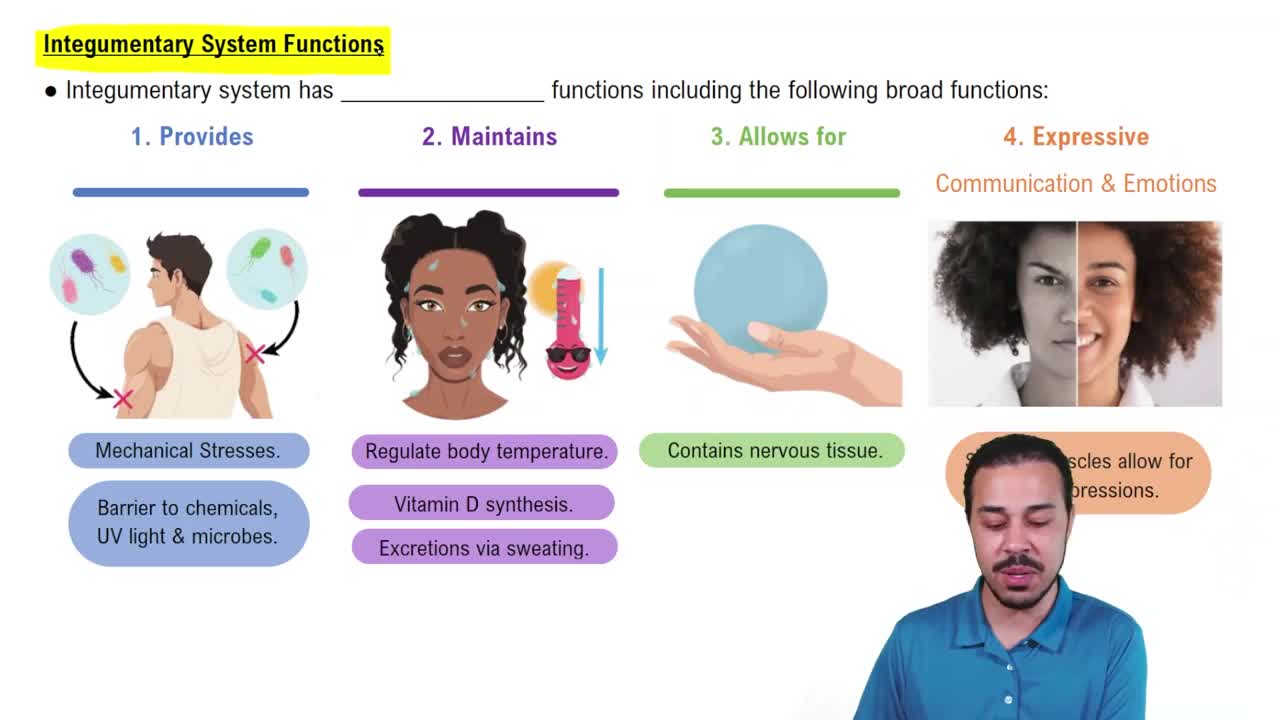
    Protection, excretion, temperature maintenance, melanin and keratin production, Vitamin D synthesis, lipid storage, and sensory detection.
  • Primary cell type in the epidermis

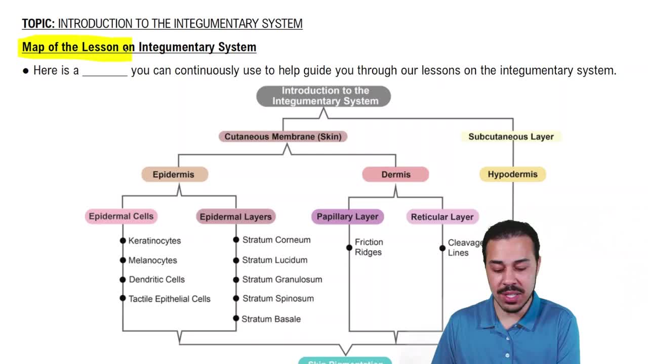
    Keratinocyte.
  • Difference between thin and thick skin

    Thin skin covers most body surface with 4 strata.
    Thick skin is on palms/soles with 5 strata including stratum lucidum.
  • Five epidermal layers from deep to superficial

    Stratum basale, stratum spinosum, stratum granulosum, stratum lucidum (thick skin only), stratum corneum.
  • Key cells/structures in the Stratum Basale

    Basal cells (stem cells), Merkel cells (touch), melanocytes (melanin), attached by hemidesmosomes.
  • Key cells/structures in the Stratum Spinosum

    8–10 layers of keratinocytes bound by desmosomes; dendritic (Langerhans) cells for immune defense.
  • What occurs in the Stratum Granulosum?

    3–5 layers of keratinocytes stop dividing, produce keratin and keratohyalin; membranes thicken and cells flatten.
  • Characteristics of the Stratum Lucidum

    Found only in thick skin; clear layer of flattened dead cells filled with keratin and keratohyalin.
  • Characteristics of the Stratum Corneum

    Outermost layer with 15–30 layers of dead keratinized cells connected by desmosomes; water resistant but not waterproof.
  • Time for cells to move from stratum basale to stratum corneum and shedding time

    Movement takes 7–10 days; cells remain on surface about 2 weeks before shedding.
  • Factors influencing skin color

    Pigments (carotene, melanin), dermal circulation (blood supply), epidermal thickness, and UV radiation exposure.
  • How do melanocytes pass pigment to keratinocytes?

    Melanin is packaged into melanosomes and transferred to keratinocytes; skin color differences depend on melanin amount and melanosome size.
  • Cause of Cyanosis

    Sustained reduction in blood oxygen causing bluish skin color, especially visible on lips and under nails.
  • Compare Basal Cell Carcinoma and Malignant Melanoma

    Basal Cell Carcinoma: common, originates in stratum basale, low metastasis, high survival.
    Malignant Melanoma: dangerous, rapid growth and metastasis, survival drops drastically if late.
  • Fiber types in the dermis and their functions

    Collagen fibers: strength and resist stretching.
    Elastic fibers: flexibility and skin recoil.
  • What are Cleavage (Tension) Lines and their clinical importance?

    Parallel collagen/elastic fiber patterns; parallel cuts heal better with less scarring, perpendicular cuts cause more scarring.
  • Cutaneous sensory receptors and their functions

    Free nerve endings: touch/pressure.
    Tactile discs: texture/steady pressure.
    Meissner's corpuscles: light touch/vibration.
    Pacinian corpuscles: deep pressure/vibration.
    Ruffini corpuscles: pressure/stretching.
  • Compare 1st, 2nd, and 3rd degree burns

    1st degree: epidermis redness.
    2nd degree: epidermis + part dermis, blistering, heals 1–2 weeks.
    3rd degree: full thickness, destroys epidermis/dermis/subcutaneous, less pain, needs grafts.
  • Purpose of the Rule of Nines

    Estimates total body surface area affected by burns for clinical assessment; modified for children.
  • Types of skin grafts based on source

    Autograft: patient's own skin.
    Allograft: cadaver skin.
    Xenograft: animal skin.
  • Difference between sebaceous glands and sebaceous follicles

    Sebaceous glands secrete sebum into hair follicles.
    Sebaceous follicles secrete sebum directly onto skin surface.
  • Compare Apocrine and Eccrine (Merocrine) sweat glands

    Apocrine: axillae, nipples, pubic; sticky, odorous secretion; hormone-influenced.
    Eccrine: all over skin, watery secretion for thermoregulation and excretion.
  • Key structures of a nail

    Nail body, lunula (pale crescent), eponychium (cuticle), hyponychium (under free edge), nail root (production site).
  • How UV radiation leads to active Vitamin D \(D_3\) (Calcitriol)

    UV converts steroid in stratum basale/spinosum to cholecalciferol (D \(3\)). Liver and kidneys convert it to calcitriol, aiding calcium/phosphate absorption.
  • Condition from Vitamin D \(3\)/Calcitriol deficiency in children

    Rickets: flexible, poorly mineralized bones due to insufficient calcium and phosphate.
  • Major age-related changes in the integumentary system

    Fewer melanocytes, drier and thinner epidermis/dermis, diminished immune response, decreased perspiration, reduced blood supply, slower repair, fewer active hair follicles.
  • Four phases of skin repair and main events

    Inflammatory: bleeding, swelling, redness.
    Migratory: scab forms, basal cells migrate, macrophages clean.
    Proliferation: epithelial cells migrate, fibroblasts synthesize collagen.
    Scarring: scab sheds, scar tissue forms.