 뒤로
Connective Tissue Types and Features in Anatomy & Physiology
 06:37

이 집합의 용어 (16)
Contains only reticular fibers (thin collagen), with reticular cells. Located in lymphoid organs like lymph nodes, bone marrow, and spleen. It forms a soft internal skeleton supporting free blood cells.
Thick collagen fibers running parallel to the direction of pull, providing very high tensile strength and flexibility.
Found in tendons, ligaments, and aponeuroses. It functions to attach structures with great strength and resist tension and stretching.
Dense irregular CT has thicker collagen bundles arranged irregularly, allowing it to withstand tension from many directions.
Located in the dermis, submucosa of the digestive tract, and fibrous capsules of organs and joints.
Similar to dense regular CT but with a very high content of elastic fibers. Found in elastic ligaments (e.g., connecting vertebrae) and walls of large arteries.
Tough but flexible tissue, avascular and without nerve fibers. Matrix secreted by chondroblasts and chondrocytes, with 80% water, collagen fibers, and sugar proteins like chondroitin and hyaluronic acid.
Hyaline cartilage, elastic cartilage, and fibrocartilage.
Inorganic calcium salts provide hardness and strength for support and protection of softer tissues.
Osteoblasts build bone, osteocytes maintain bone, and osteoclasts break down bone.
Bone is richly vascularized and contains more collagen than cartilage.
Because it consists of cells suspended in a nonliving fluid matrix (blood plasma), with soluble protein fibers visible only during clotting.
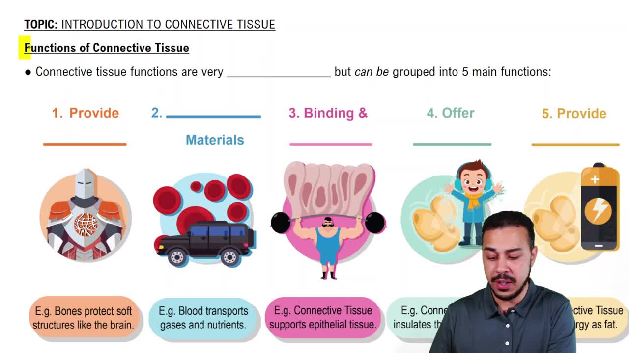
Transporting nutrients, wastes, gases, and other substances throughout the body.
Collagen fibers provide high tensile strength.
Osteoblasts are the cells that form bone.
Areolar connective tissue soaks up fluid when edema occurs.