 뒤로
Muscle Anatomy & Physiology Flashcards
 6:31

이 집합의 용어 (29)
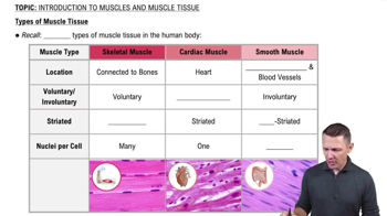
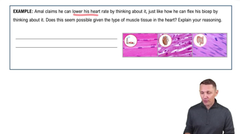
Skeletal, Cardiac, and Smooth muscle.
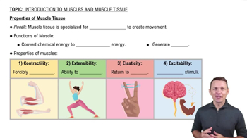
Excitability: respond to stimuli; Contractility: shorten when stimulated; Extensibility: stretchable; Elasticity: recoil to resting length.
Epimysium, made of dense irregular connective tissue.
Perimysium, made of fibrous connective tissue.
Endomysium, made of fine areolar connective tissue.
The plasma membrane of a muscle fiber (cell).
Elongated, cylindrical cell with multiple peripheral nuclei, many mitochondria, glycosomes, myoglobin, myofibrils, sarcoplasmic reticulum, and T tubules.
Densely packed, rodlike contractile elements making up about 80% of muscle cell volume, showing striations.
The region of a myofibril between two successive Z discs, the smallest contractile unit.
Thick filaments are made of myosin; thin filaments are made of actin.
Z-discs anchor thin filaments and connect adjacent myofibrils.
The lighter region in the center of the A band where thick and thin filaments do not overlap.
The protein myomesin line that holds adjacent thick filaments together in the sarcomere center.
One site binds to actin and the other binds to ATP.
Tropomyosin blocks myosin binding sites on actin at rest, regulating contraction.
TnI: inhibits contraction by binding actin; TnT: binds tropomyosin; TnC: binds calcium ions.
Regulates intracellular calcium ion levels essential for contraction.
A structure formed by a T-tubule flanked by two terminal cisternae of the SR.
T-tubules conduct action potentials deep into the fiber, triggering SR to release calcium from terminal cisternae for contraction.
Shortening means sarcomeres shorten as thin filaments slide past thick filaments during contraction.
Myosin heads bind to actin, pull thin filaments toward the M line, shortening sarcomeres and muscle fibers.
The synapse between a motor neuron axon terminal and a skeletal muscle fiber's motor end plate.
Acetylcholine (ACh) is released to activate contraction; acetylcholinesterase breaks down ACh to stop contraction.
Low Ca2+: tropomyosin blocks actin sites, muscle relaxes. High Ca2+: Ca2+ binds troponin, moves tropomyosin, allowing cross-bridge cycling.
Action potential reaches motor neuron terminal → Ca2+ influx → ACh release → ACh binds receptors → Na+ influx depolarizes muscle membrane.
Action potential spreads along sarcolemma and T-tubules → triggers Ca2+ release from SR → initiates contraction.
1. Cross-bridge formation (myosin binds actin); 2. Power stroke (myosin pulls actin); 3. Cross-bridge detachment (ATP binds myosin); 4. Cocking of myosin head (ATP hydrolysis).
A motor neuron and all the muscle fibers it innervates, contracting together.
Latent period (excitation-contraction coupling), contraction period (cross-bridge cycling), relaxation period (Ca2+ reuptake).