 뒤로
Muscle Tissue and Contraction - Anatomy & Physiology
 00:36

이 집합의 용어 (28)
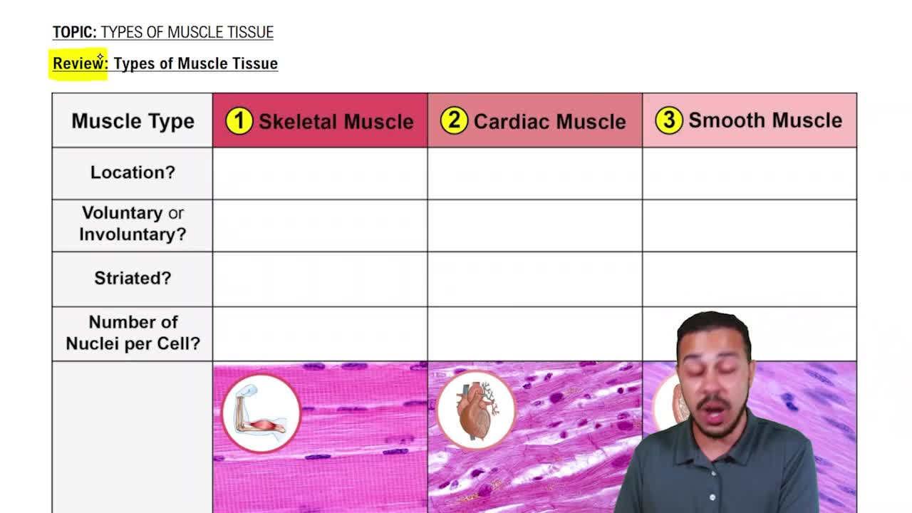
The three types are skeletal, cardiac, and smooth muscle.
Elongated muscle cells found in skeletal and smooth muscle, but not in cardiac muscle.
Sarcolemma is the plasma membrane of muscle cells; sarcoplasm is the cytoplasm of muscle cells.
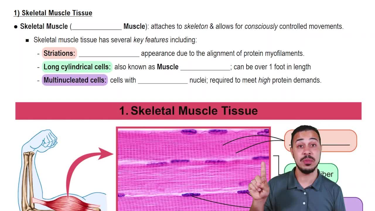
Skeletal muscle is striated, voluntary, and responsible for body mobility.
Cardiac muscle is striated, involuntary, and found only in the heart.
Smooth muscle is nonstriated, involuntary, and found in walls of hollow organs.
Excitability, contractility, extensibility, and elasticity.
Produce movement, maintain posture, stabilize joints, and generate heat.
Epimysium surrounds whole muscle, perimysium surrounds fascicles, endomysium surrounds individual fibers.
Direct attachments fuse epimysium to bone periosteum; indirect use tendons or aponeuroses.
Fibers are long cylindrical cells, 10-100 μm diameter, up to 30 cm long, with multiple nuclei beneath sarcolemma.
Contains glycosomes (glycogen storage) and myoglobin (oxygen storage).
Rodlike structures in muscle fibers made of sarcomeres; account for ~80% of cell volume.
Alternating dark A bands and light I bands aligned along myofibrils.
Smallest contractile unit of muscle fiber, between two Z discs, about 2 μm long.
Thick filaments contain myosin; thin filaments contain actin, tropomyosin, and troponin.
Titin is an elastic filament that stabilizes thick filaments and helps muscle recoil after stretching.
Dystrophin links thin filaments to sarcolemma proteins, stabilizing muscle fiber structure.
Specialized smooth ER that stores and releases calcium ions to trigger muscle contraction.
Invaginations of sarcolemma that conduct action potentials deep into muscle fibers, triggering calcium release.
Structure formed by a T tubule flanked by two terminal cisterns of the SR, coordinating contraction signals.
During contraction, thin filaments slide past thick filaments, shortening sarcomeres without changing filament length.
I bands and H zones shorten; A bands length stays the same; Z discs move closer.
Sequence where an action potential triggers calcium release, leading to myofilament sliding and muscle contraction.
Calcium binds to troponin, causing tropomyosin to move and expose myosin-binding sites on actin.
Myosin heads bind actin, pivot, detach using ATP, and reattach to pull thin filaments during contraction.
After death, ATP depletion prevents cross bridge detachment, causing muscle stiffness.
Genetic disease caused by defective dystrophin, leading to muscle degeneration and weakness.