 뒤로
Muscle Tissue and Physiology - Anatomy & Physiology
 6:31

이 집합의 용어 (31)
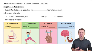
the ability of muscle cells to shorten forcefully, generating tension.
the ability of muscle cells to respond to stimuli by depolarizing their membrane.
the ability of muscle cells to propagate an electrical signal along their membrane.
the ability of muscle cells to stretch without damage.
the ability of muscle cells to return to their original length after stretching.
a muscle cell, also called a muscle fiber in skeletal muscle.
the cytoplasm of a muscle cell, containing organelles and myofibrils.
the plasma membrane of a muscle cell that conducts action potentials.
a cylindrical organelle composed of contractile proteins within muscle cells.
stores and releases calcium ions to trigger muscle contraction.
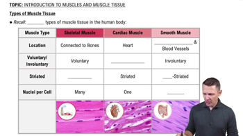
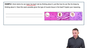
Skeletal muscle is voluntary, striated, and attached to bones; cardiac muscle is involuntary, striated, and found in the heart; smooth muscle is involuntary, non-striated, and found in organs.
invaginations of the sarcolemma that transmit action potentials deep into muscle fibers.
contractile proteins actin (thin filament) and myosin (thick filament) within myofibrils.
the functional contractile unit of a myofibril, defined by Z discs.
describes how actin and myosin filaments slide past each other to shorten sarcomeres during contraction.
regulates intracellular calcium levels by storing and releasing calcium ions.
conduct action potentials from the sarcolemma to the sarcoplasmic reticulum to trigger calcium release.
A triad consists of one T-tubule flanked by two terminal cisternae of the sarcoplasmic reticulum.
Contractile proteins (actin, myosin) generate tension; regulatory proteins (troponin, tropomyosin) control contraction; structural proteins (titin) maintain filament alignment.
The myosin head has an actin binding site for crossbridge formation and a myosin ATPase site for ATP hydrolysis.
the connection formed when the myosin head binds to the active site on actin during contraction.
At low calcium, tropomyosin blocks actin's active sites, and troponin holds tropomyosin in place, preventing crossbridge formation.
At high calcium, calcium binds to troponin, causing tropomyosin to move and expose actin's active sites for crossbridge formation.
Membrane potential is the voltage difference across a cell membrane due to ion distribution.
the stable, negative membrane potential of a resting cell, typically around -70 mV in muscle cells.
reduction of membrane potential as sodium ions enters the cell, making it more positive.
the restoration of the negative membrane potential as potassium ions exit the cell.
The sodium-potassium pump uses ATP to move 3 sodium ions out and 2 potassium ions into the cell, maintaining ion gradients.
The neuromuscular junction includes the motor neuron axon terminal, synaptic cleft, and motor end plate of the muscle fiber.
ACh is the neurotransmitter that binds receptors on the motor end plate to initiate muscle cell depolarization.
Acetylcholinesterase breaks down ACh in the synaptic cleft to terminate the signal and allow muscle relaxation.