뒤로

Muscle Tissue and Skeletal Muscle Structure - Anatomy & Physiology

컨트롤 버튼이 '내비게이션' 모드로 변경되었습니다.
1/20
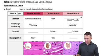
  • Three types of muscle tissue

    Skeletal: attached to bones/skin, striated, voluntary.
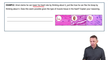
    Cardiac: heart walls, striated, involuntary.
    Smooth: walls of hollow organs, non-striated, involuntary.
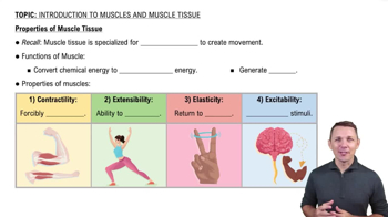
  • Special characteristics of muscle tissue

    Excitability: respond to stimuli.
    Contractility: shorten when stimulated.
    Extensibility: stretchable.
    Elasticity: recoil to resting length.
  • Functions of muscles

    Movement of bones, circulation of body fluids, maintaining posture, stabilizing joints, and heat generation.
  • Connective tissue coverings of skeletal muscle

    Epimysium: surrounds entire muscle.
    Perimysium: surrounds fascicles.
    Endomysium: surrounds individual muscle fibers.
  • Structure of a skeletal muscle fiber

    Cylindrical cell with multiple peripheral nuclei, many mitochondria, containing myofibrils, sarcoplasmic reticulum, and T tubules.
  • Myofibrils

    Rodlike elements inside muscle fibers that exhibit striations and contain sarcomeres, the contractile units.
  • Sarcomere

    Smallest contractile unit of muscle fiber, between two Z discs, composed of thick and thin filaments.
  • Thick filaments

    Composed of myosin protein with heads that bind actin and ATP during contraction.
  • Thin filaments

    Composed of actin, with regulatory proteins tropomyosin and troponin controlling myosin binding sites.
  • Sarcoplasmic reticulum (SR)

    Smooth ER surrounding myofibrils, stores and regulates intracellular Ca2+.
  • T tubules

    Invaginations of sarcolemma that penetrate muscle fiber and associate with SR to form triads.
  • Sliding filament model of contraction

    Myosin heads bind, pull thin filaments toward M line, shortening sarcomeres and muscle fibers.
  • Neuromuscular junction

    Synapse where motor neuron axon terminal meets muscle fiber sarcolemma, releasing acetylcholine (ACh).
  • Events at the neuromuscular junction

    Action potential triggers Ca2+ entry, ACh release, ACh binds receptors, ion channels open, generating muscle action potential.
  • Generation of muscle action potential

    Local depolarization by Na+ influx, if threshold reached, voltage-gated Na+ channels open, propagating action potential.
  • Repolarization phase

    Na+ channels close, K+ channels open, K+ efflux restores resting membrane potential.
  • Role of Ca2+ in muscle contraction

    Ca2+ binds troponin, moves tropomyosin, exposing actin active sites for myosin binding.
  • Cross bridge cycle steps

    1. Myosin head binds actin.
    2. Power stroke pulls thin filament.
    3. ATP binds, cross bridge detaches.
    4. ATP hydrolysis cocks myosin head.
  • Muscle fiber relaxation

    Ca2+ pumped back into SR, tropomyosin blocks actin sites, myosin heads detach, muscle relaxes.
  • Organization levels of skeletal muscle

    Muscle > fascicle > muscle fiber > myofibril > sarcomere.