Skip to main content
Anatomy & Physiology
나의 코스
배우기
시험 준비
AI 튜터
학습 가이드
교과서적 해결책
플래시카드
탐험해 보세요
앱을 사용해 보세요
나의 코스
배우기
시험 준비
AI 튜터
학습 가이드
교과서적 해결책
플래시카드
탐험해 보세요
앱을 사용해 보세요
뒤로
Muscle Tissue Structure and Function - Anatomy & Physiology
카드를 뒤집기 위해 탭할 수 있습니다.
Layers of connective tissue in skeletal muscle
카드를 뒤집기 위해 탭할 수 있습니다.
👆
Layers of connective tissue in skeletal muscle
Epimysium
surrounds entire muscle,
Perimysium
surrounds fascicles,
Endomysium
surrounds individual muscle fibers.
진행 상황 추적
컨트롤 버튼이 '내비게이션' 모드로 변경되었습니다.
1/20
추천 영상
VideoThumbView.guidedCourse
6:31
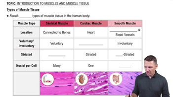
Types of Muscle Tissue
18731
views
494
rank
1
comments
VideoThumbView.guidedCourse
2:00
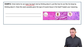
Introduction to Muscles and Muscle Tissue Example 1
16655
views
324
rank
VideoThumbView.guidedCourse
4:36
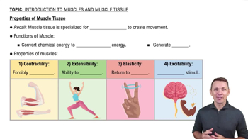
Properties of Muscle Tissue
25637
views
741
rank
2
comments
이 집합의 용어 (20)
하이드의 정의
Layers of connective tissue in skeletal muscle
Epimysium
surrounds entire muscle,
Perimysium
surrounds fascicles,
Endomysium
surrounds individual muscle fibers.
Definition of a muscle fascicle
A bundle of muscle fibers surrounded by
perimysium
connective tissue.
Structure of a muscle fiber
A muscle fiber is a single muscle cell, surrounded by
sarcolemma
, containing many
myofibrils
.
Function of the sarcolemma
The muscle cell membrane that encloses the sarcoplasm and conducts action potentials.
Role of myofibrils in muscle fibers
Myofibrils are bundles of protein filaments responsible for muscle contraction.
Basic contractile unit of muscle
The
sarcomere
, which extends from one Z line to the next.
Components of a sarcomere
Contains thick filaments (myosin), thin filaments (actin), Z lines, M line, H zone, I band, and A band.
Function of the Z line in sarcomeres
Defines the boundary between adjacent sarcomeres and anchors thin filaments.
Difference between A band and I band
A band contains thick filaments and overlapping thin filaments; I band contains only thin filaments.
Role of the H zone
Central region of the A band with only thick filaments, no thin filament overlap.
Structure of thick filaments
Composed mainly of myosin molecules with heads that form cross-bridges during contraction.
Structure of thin filaments
Made of F actin strands, tropomyosin, troponin complex, and nebulin.
Function of tropomyosin and troponin
Tropomyosin blocks active sites on actin; troponin binds calcium to move tropomyosin and expose active sites.
Sliding filament model of contraction
Myosin heads bind to actin active sites, pivot, and slide thin filaments past thick filaments to shorten sarcomeres.
Role of calcium ions in muscle contraction
Ca2+ binds to troponin, causing tropomyosin to move and expose actin active sites for myosin binding.
Function of transverse (T) tubules
Transmit action potentials deep into muscle fibers to trigger calcium release from the sarcoplasmic reticulum.
Role of sarcoplasmic reticulum in muscle fibers
Stores and releases Ca2+ to initiate muscle contraction.
Definition of a motor unit
A motor neuron and all the muscle fibers it innervates, contracting together.
All-or-none principle in muscle contraction
Individual muscle fibers contract fully or not at all when stimulated.
Difference between isotonic and isometric contractions
Isotonic changes muscle length with constant tension; isometric develops tension without changing length.
 뒤로뒤로
 6:31