 뒤로
Renal Anatomy and Physiology

 0:54

이 집합의 용어 (20)
A nephron consists of the renal corpuscle (glomerulus and Bowman's capsule) and the renal tubule (proximal tubule, loop of Henle, distal tubule, and collecting duct).
Filtration-Blood pressure substances from glomerular capillaries into Bowman’s capsule/reabsorption-Essential substances reclaimed from renal tubule moved into peritubular capillaries/secretion Additional waste products moved from blood to renal tubules /are the key processes in urine formation.
Glomerular hydrostatic pressure promotes filtration by pushing fluid out of capillaries.
Capsular hydrostatic pressure and blood colloid osmotic pressure oppose filtration by pushing fluid back into capillaries.
Includes myogenic mechanism and tubuloglomerular feedback to maintain stable GFR despite blood pressure changes.
Involves sympathetic nervous system and renin-angiotensin system to regulate GFR during stress or low blood volume.
Reabsorption occurs via active transport, osmosis, and diffusion from renal tubules into peritubular capillaries.
Controlled by hormones like aldosterone (sodium) and antidiuretic hormone (ADH) (water).
Tubular secretion removes substances like H+, potassium, and drugs from blood into tubules for excretion.
Occurs when ADH is low, allowing water to remain in tubules and be excreted, producing dilute urine.
Occurs when ADH is high, increasing water reabsorption and concentrating urine.
Urine is typically clear, pale yellow, with a specific gravity of about 1.003 to 1.030.
Contains mostly water, urea, creatinine, and electrolytes.
Glucose (diabetes mellitus), protein (kidney damage), ketones (starvation or diabetes), and blood (infection or trauma).
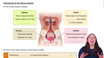
Ureters connect kidneys to bladder; bladder stores urine; urethra conducts urine out of the body.
Muscular tubes with smooth muscle layers that propel urine by peristalsis from kidneys to bladder.
Hollow, muscular organ with a transitional epithelium lining and detrusor muscle for urine storage and expulsion.
Tube lined with mucosa and smooth muscle that carries urine from bladder to exterior.
Micturition is the process of urine voiding from the bladder.
Involves parasympathetic stimulation to contract bladder and somatic control of external sphincter for voluntary urination.