Skip to main content
Anatomy & Physiology
나의 코스
배우기
시험 준비
AI 튜터
학습 가이드
교과서적 해결책
플래시카드
탐험해 보세요
앱을 사용해 보세요
나의 코스
배우기
시험 준비
AI 튜터
학습 가이드
교과서적 해결책
플래시카드
탐험해 보세요
앱을 사용해 보세요
뒤로
Skeletal Muscle Tissue Structure and Contraction
카드를 뒤집기 위해 탭할 수 있습니다.
What are the main functions of skeletal muscle tissue?
카드를 뒤집기 위해 탭할 수 있습니다.
👆
What are the main functions of skeletal muscle tissue?
Movement
,
posture maintenance
,
body temperature regulation
, and
guarding body entrances/exits
(sphincters).
진행 상황 추적
컨트롤 버튼이 '내비게이션' 모드로 변경되었습니다.
1/20
추천 영상
VideoThumbView.guidedCourse
6:31
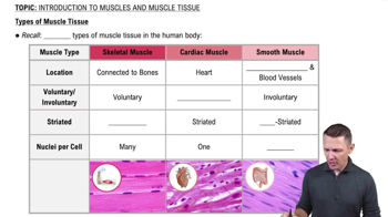
Types of Muscle Tissue
18703
views
494
rank
1
comments
VideoThumbView.guidedCourse
2:00
Introduction to Muscles and Muscle Tissue Example 1
16631
views
323
rank
VideoThumbView.guidedCourse
4:36
Properties of Muscle Tissue
25596
views
741
rank
2
comments
이 집합의 용어 (20)
하이드의 정의
What are the main functions of skeletal muscle tissue?
Movement
,
posture maintenance
,
body temperature regulation
, and
guarding body entrances/exits
(sphincters).
Describe the general structure of a skeletal muscle fiber.
Long, cylindrical, multinucleated cells with striations, containing myofibrils made of thin (actin) and thick (myosin) filaments.
What is a sarcomere and what are its key components?
The sarcomere is the functional contractile unit of muscle, composed of
thin filaments
,
thick filaments
,
Z lines
,
M line
,
A band
,
I band
, and
H zone
.
What changes occur in the sarcomere during muscle contraction?
The
H zone
and
I band
shorten, the
A band
remains the same length, and thin filaments slide over thick filaments.
What role does the neuromuscular junction play in muscle contraction?
It transmits the nerve impulse to the muscle fiber, releasing acetylcholine (ACh) to trigger an action potential.
How does acetylcholine (ACh) initiate muscle contraction?
ACh binds to receptors on the sarcolemma, opening Na+ channels and generating an action potential.
What happens to acetylcholine after it triggers an action potential?
It is broken down by acetylcholinesterase (AChE) to stop the signal and allow muscle relaxation.
Describe the role of calcium ions (Ca2+) in muscle contraction.
Ca2+ binds to troponin, causing tropomyosin to move and expose active sites on actin for myosin binding.
What is the function of troponin and tropomyosin in muscle fibers?
Troponin binds Ca2+ to move tropomyosin, which blocks or exposes active sites on actin for contraction control.
Explain the cross-bridge cycle in muscle contraction.
Myosin heads bind to actin active sites, pivot to pull filaments (power stroke), detach with ATP, and reactivate for another cycle.
What is the source of energy for muscle contraction?
ATP hydrolysis provides energy for myosin head movement and detachment during the contraction cycle.
How do muscle fibers develop during growth?
Muscle fibers form by fusion of myoblasts into multinucleated myotubes that mature into muscle fibers.
What is the role of the sarcoplasmic reticulum in muscle contraction?
It stores and releases Ca2+ ions in response to action potentials to regulate contraction.
What is the function of T tubules in skeletal muscle fibers?
T tubules transmit action potentials deep into the muscle fiber to trigger Ca2+ release from the sarcoplasmic reticulum.
What is Duchenne Muscular Dystrophy (DMD)?
A severe X-linked recessive disorder causing muscle wasting due to absence of dystrophin, leading to muscle damage and weakness.
How does the absence of dystrophin affect muscle fibers in DMD?
Without dystrophin, muscle fibers are damaged during contraction, causing progressive muscle degeneration.
What is the typical prognosis for individuals with Duchenne Muscular Dystrophy?
Most become wheelchair-dependent by age 12, with median life expectancy of 20-30 years due to cardiac or respiratory failure.
What structural features distinguish skeletal, cardiac, and smooth muscle tissues?
Skeletal: long, cylindrical, multinucleated, striated; Cardiac: short, branched, striated, intercalated discs; Smooth: short, spindle-shaped, nonstriated.
What is the role of titin in the sarcomere?
Titin stabilizes thick filaments and provides elasticity, helping sarcomeres return to resting length after contraction.
What happens during the power stroke in muscle contraction?
Myosin heads pivot, pulling thin filaments toward the M line, shortening the sarcomere and generating tension.
 뒤로뒤로
 6:31