 뒤로
Tissue and Skin Microscopy in Anatomy & Physiology
 03:32

이 집합의 용어 (20)
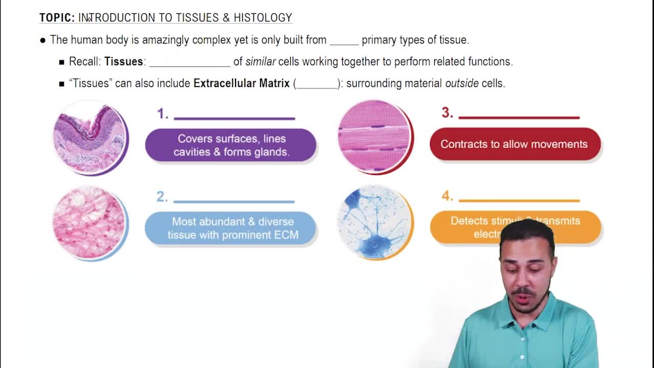
Epithelial tissue covers body surfaces and lines cavities, providing protection, absorption, secretion, and sensation.
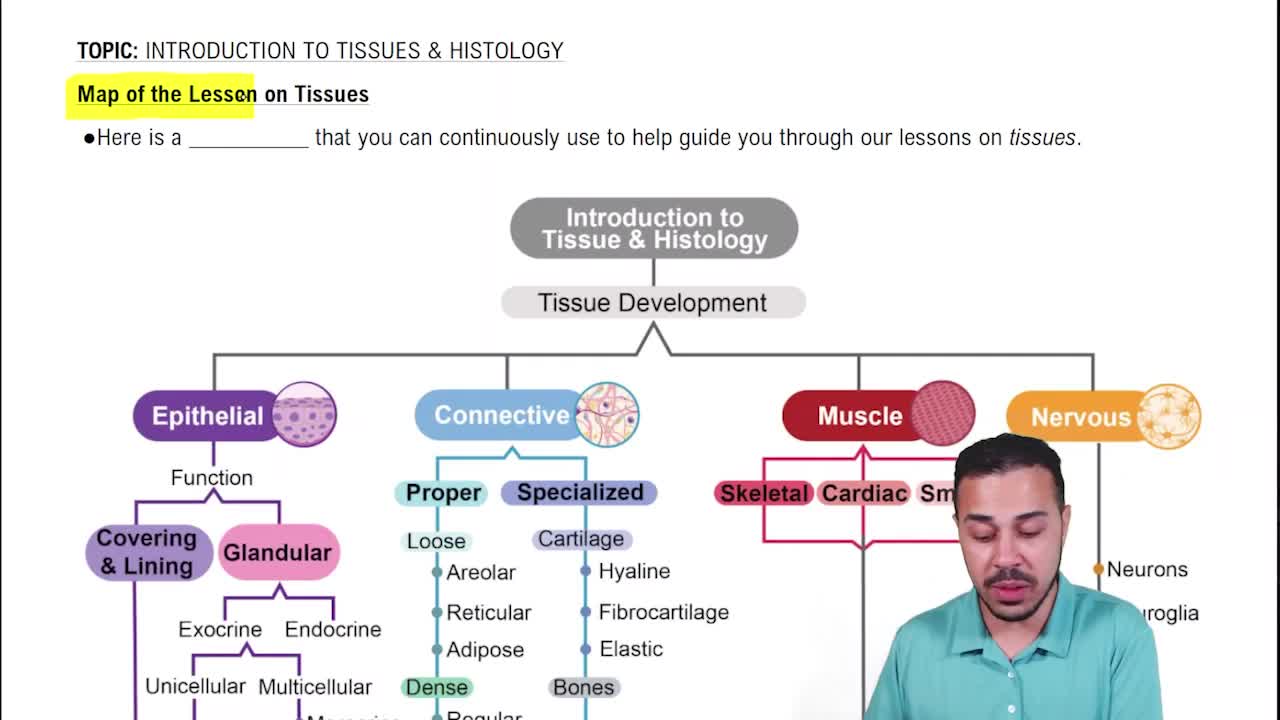
Epithelial, connective, muscle, and nervous tissues.
It has abundant extracellular matrix with scattered cells, providing support and binding other tissues.
Muscle tissue shows elongated cells called fibers, often striated in skeletal muscle, and specialized for contraction.
Nervous tissue contains neurons with large cell bodies and long processes, plus supporting glial cells.
The epidermis, dermis, and sometimes the hypodermis (subcutaneous layer).
Keratinized stratified squamous epithelium, which protects against abrasion and water loss.
Connective tissue, blood vessels, nerves, hair follicles, and glands.
It contains loose connective tissue and abundant adipose tissue for insulation and cushioning.
They produce keratin, a protein that strengthens the skin and makes it waterproof.
Coiled tubular structures lined with cuboidal or columnar epithelial cells in the dermis.
They produce melanin, the pigment that protects against UV radiation.
Dense irregular connective tissue, providing strength and elasticity.
Thick skin has a thicker epidermis with a prominent stratum corneum and no hair follicles; thin skin has hair follicles and a thinner epidermis.
The deepest layer of the epidermis where basal cells divide to produce new keratinocytes.
The outermost layer of dead keratinized cells that forms a tough protective barrier.
As small lumens lined by endothelial cells within the dermis.
Invaginations of the epidermis into the dermis surrounding a hair shaft.
They are immune cells in the epidermis that detect and present antigens.
Large, empty-looking cells with thin cytoplasm and nuclei pushed to the side, storing fat.