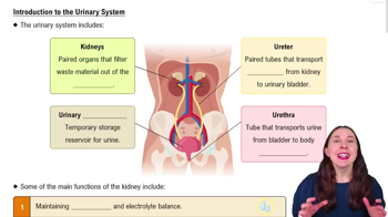
Regulate total water volume and solute concentration, maintain ECF ion concentrations, ensure acid-base balance, and remove metabolic wastes, toxins, and drugs.
Endocrine functions of the kidneys
Secrete renin for blood pressure regulation, erythropoietin for RBC production, activate vitamin D, and perform gluconeogenesis during prolonged fasting.
Organs of the urinary system
Includes kidneys (major excretory organs), ureters (transport urine to bladder), urinary bladder (temporary urine storage), and urethra (urine excretion).
Location of the kidneys
Located against the posterior body wall, typically at the level of the 12th rib, retroperitoneal with supportive layers including renal fascia, perirenal fat, and fibrous capsule.
Internal anatomy of the kidney
Consists of the renal cortex, renal medulla (with pyramids), renal pelvis, minor and major calyces, and the renal hilum where vessels and ureter enter/exit.
Blood supply to the kidneys
Renal arteries deliver ~25% of cardiac output; blood flows through segmental, interlobar, arcuate, and cortical radiate arteries to the nephrons.
Nephron structure
Functional unit of the kidney with two main parts: renal corpuscle (glomerulus and Bowman's capsule) and renal tubule (proximal tubule, loop of Henle, distal tubule).
Function of the glomerulus
A tuft of fenestrated capillaries specialized for filtration of blood plasma into the Bowman's capsule.
Bowman's capsule layers
Has a parietal layer (outer) and a visceral layer with podocytes that wrap around glomerular capillaries.
Classes of nephrons
Cortical nephrons (85%, mostly in cortex) and juxtamedullary nephrons (long loops into medulla, important for concentrated urine).
Juxtaglomerular complex (JGC) components
Includes macula densa cells (sense NaCl), granular cells (secrete renin), and extraglomerular mesangial cells (signal transmission).
Role of granular cells in JGC
Mechanoreceptors that sense blood pressure in afferent arteriole and secrete renin to regulate blood pressure.
Urine physical characteristics
Normally clear and pale to deep yellow due to urochrome; cloudy urine may indicate infection; odor can change with standing or diet.
Urine pH and specific gravity
Slightly acidic (~pH 6, range 4.5-8.0); specific gravity ranges from 1.001 to 1.035 depending on solute concentration.
Chemical composition of urine
About 95% water and 5% solutes including nitrogenous wastes like urea, uric acid, creatinine, and ions such as Na+, K+, PO4 3-.
Ureter anatomy and function
Retroperitoneal tubes that convey urine from kidneys to bladder; distal ends close during bladder contraction to prevent backflow.
Layers of the ureter wall
Includes mucosa with transitional epithelium, muscularis with longitudinal and circular layers, and outer adventitia.
Urinary bladder anatomy
Muscular sac for urine storage, located retroperitoneally on pelvic floor; has openings for ureters and urethra forming the trigone.
Bladder capacity and features
Can hold ~500 ml normally, expandable to twice that; collapses when empty with rugae; can burst if overdistended.
Urethra structure and sphincters
Muscular tube lined mostly by pseudostratified columnar epithelium; internal sphincter is involuntary smooth muscle, external sphincter is voluntary skeletal muscle.
 뒤로

 0:54