 뒤로
General Biology: Cell Structure, Membranes, and Signaling
 04:26

이 집합의 용어 (28)
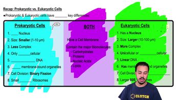
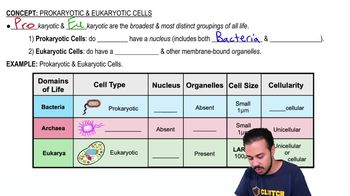
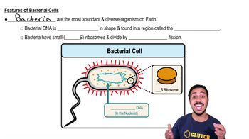
Prokaryotes lack a membrane-bound nucleus and organelles; DNA is in a nucleoid. Eukaryotes have a nucleus and membrane-bound organelles. Bacteria have peptidoglycan cell walls; archaea and eukaryotes do not.
Surface area increases with the square of cell length, volume with the cube. Larger cells have a lower surface-area-to-volume ratio, reducing exchange efficiency.
The nuclear envelope separates nucleus from cytoplasm with two membranes; nuclear pores regulate RNA, protein, and ribosomal subunit movement.
The nucleolus synthesizes and processes ribosomal RNA and assembles ribosomal subunits for export through nuclear pores.
Free ribosomes make proteins for the cytosol; bound ribosomes on rough ER make proteins for secretion or membranes. Targeting depends on protein signals, not ribosome type.
They have their own DNA, bacterial-like ribosomes, divide by binary fission, and have double membranes, indicating bacterial ancestry.
Includes nuclear envelope, ER, Golgi apparatus, endosomes, lysosomes, vesicles, vacuoles, and plasma membrane involved in membrane trafficking.
Rough ER synthesizes and processes secreted and membrane proteins; smooth ER synthesizes lipids, detoxifies, and stores calcium.
Ribosome on rough ER → ER lumen → ER exit vesicles → Golgi cis face → Golgi trans face → secretory vesicles → exocytosis.
Lysosomes contain acid hydrolases that digest macromolecules and worn cellular components; defects cause substrate accumulation (e.g., Tay–Sachs disease).
Microtubules support organelle transport, spindle formation, and cilia/flagella movement; dynamic assembly/disassembly moves chromosomes during division.
Actin filaments maintain cell shape, enable muscle contraction with myosin, cytoplasmic streaming, and form the contractile ring in cytokinesis.
Contain a 9 + 2 microtubule arrangement; dynein motors use ATP to slide microtubules, causing bending; basal bodies anchor them.
Membranes are phospholipid bilayers with embedded proteins that move laterally; hydrophilic heads face out, hydrophobic tails face in.
Unsaturated fatty acids increase fluidity; saturated tails decrease it. Cholesterol buffers fluidity by restraining movement at high temps and preventing solidification at low temps.
Simple diffusion passes small nonpolar molecules through the bilayer; facilitated diffusion uses transport proteins for ions and polar molecules without energy.
Primary uses ATP directly to move substances against gradients; secondary uses downhill movement of one solute to drive uphill transport of another.
Moves 3 Na⁺ out and 2 K⁺ in per ATP hydrolyzed, creating an electrogenic effect contributing to membrane potential.
Phagocytosis engulfs large particles; pinocytosis internalizes fluid nonselectively; receptor-mediated endocytosis selectively concentrates cargo via receptors.
Reception: ligand binds receptor; transduction: signal relayed inside cell; response: cellular activity changes.
Ligand binding causes GDP-GTP exchange on G protein, activating it to regulate effectors; GTP hydrolysis returns it to inactive state.
Ligand binding induces receptor dimerization and tyrosine phosphorylation, recruiting proteins to initiate downstream signaling.
Small molecules like cAMP, Ca²⁺, IP₃ relay and amplify signals inside the cell from activated receptors.
IP₃ opens ER calcium channels, releasing Ca²⁺ into cytosol; Ca²⁺ and diacylglycerol activate protein kinase C and other targets.
Protein kinases add phosphate groups to activate or inhibit proteins; phosphatases remove them to reset signaling.
Integral proteins embed in the bilayer; transmembrane proteins span it; peripheral proteins associate with membrane surfaces.
Carbohydrate chains on glycoproteins and glycolipids project outward and aid in cell recognition.
They increase surface area relative to volume, enhancing nutrient uptake and waste removal efficiency.