Skip to main content
General Biology
나의 코스
배우기
시험 준비
AI 튜터
학습 가이드
교과서적 해결책
플래시카드
탐험해 보세요
앱을 사용해 보세요
나의 코스
배우기
시험 준비
AI 튜터
학습 가이드
교과서적 해결책
플래시카드
탐험해 보세요
앱을 사용해 보세요
뒤로
Microscopy and Cytology - General Biology lab 5
카드를 뒤집기 위해 탭할 수 있습니다.
Function of the arm on a compound microscope
카드를 뒤집기 위해 탭할 수 있습니다.
👆
Function of the arm on a compound microscope
Used to carry the microscope
; located between the body tube and the base.
진행 상황 추적
컨트롤 버튼이 '내비게이션' 모드로 변경되었습니다.
1/20
추천 영상
06:35
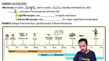
Microscopes
24938
views
567
rank
02:52
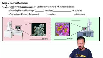
Types of Electron Microscopes
17891
views
357
rank
1
comments
이 집합의 용어 (20)
하이드의 정의
Function of the arm on a compound microscope
Used to carry the microscope
; located between the body tube and the base.
Purpose of the condenser in a compound microscope
Substage lens that concentrates light on the specimen
; its position can be adjusted to vary light intensity.
What is total magnification in microscopy?
The product of the ocular lens magnification and the objective lens magnification.
Magnification of the ocular lens in the lab microscopes
10X
; the eyepiece magnifies the image ten times.
Magnifications of the four objective lenses used
Scanning: 4X, Low power: 10X, High power: 40X, Oil immersion: 100X.
How to calculate total magnification with a 40X objective lens
Total magnification = 10X (ocular) × 40X (objective) = \(400X\).
Definition of resolution in microscopy
Resolving power
is the ability to distinguish two close objects as separate; compound microscopes resolve objects about 0.2 µm apart.
What is parfocal in compound microscopes?
Once focused on low power, the image remains nearly in focus when switching to higher magnifications.
Proper use of coarse and fine focus knobs
Use coarse focus only on low power; use fine focus for final adjustments and on high power.
Function of the iris diaphragm
Regulates the amount of light passing through the stage to improve image contrast.
What is the field of view in microscopy?
The circular area visible through the microscope lens; its diameter decreases with increasing magnification.
What is depth of field in microscopy?
The vertical distance in focus; decreases as magnification increases.
How to prepare a wet mount slide
Place specimen on slide, add liquid (water or stain), cover with coverslip avoiding air bubbles, then observe under microscope.
Purpose of staining specimens
Stains absorb light and improve contrast to reveal cellular structures.
Ocular micrometer calibration at low power (10X objective)
One ocular unit equals \(10\ \mu m\).
Ocular micrometer calibration at high power (40X objective)
One ocular unit equals \(2.5\ \mu m\).
How to measure specimen size using an ocular micrometer
Count ocular units spanning the specimen and multiply by the ocular unit length for the objective used.
Difference between red and white blood cells under microscope
Red blood cells are small, round, pinkish, and lack a nucleus; white blood cells are larger with dark-stained nuclei.
Why never use coarse focus on high power
Coarse focus can damage the slide or lens because the objective is very close to the specimen.
How to carry a microscope safely
Use both hands: one on the arm and one under the base; keep the cord secure to avoid tripping.
 뒤로뒤로
 06:35