What are the main optical components of a light microscope?
The main optical components are the condenser lens, objective lens, and ocular lens.
What is the role of the condenser lens in a light microscope?
The condenser lens projects a cone of light to illuminate the specimen but does not magnify the image.
How is total magnification calculated in a light microscope?
Total magnification is the product of the magnifying power of the objective lens and the ocular lens.
Define resolution in microscopy.
Resolution is the smallest distance between two points that can be distinguished as separate entities.
Why is resolution more important than magnification for image quality?
High magnification without high resolution results in blurred images with little detail.
What is the typical resolving power of the best light microscopes?
The resolving power is about 0.2 micrometers (µm).
What does the numerical aperture (NA) of a lens represent?
NA is a function of the lens's light-collecting ability and affects the resolution and brightness of the image.
Write the formula for resolution in microscopy.
Resolution = \(\frac{0.61 \lambda}{n \sin \theta}\), where λ is wavelength, n is refractive index, and θ is half the angular width of the cone of light.
What precautions should be taken when handling microscope objectives?
Never touch the glass part of objectives and use the course adjustment knob only with 4x objective.
What is the function of the coarse and fine focus knobs?
The coarse focus knob moves the stage quickly for rough focusing; the fine focus knob adjusts focus precisely.
What does it mean that microscopes are parfocal?
If one objective lens is in focus, the others will be close to focus when rotated into position.
What does it mean that microscopes are parcentral?
An object centered in the field of view with one lens will be close to center with other lenses.
Describe the general shape and features of squamous epithelial cells from the oral mucosa.
They are flattened, polygonal cells with distinct plasma membranes, finely granular cytoplasm, and centrally or eccentrically placed small rounded or oval nuclei.
What stain is used to observe cheek cells in this lab?
Methylene Blue stain is used to color the cheek cells for better visualization.
What are the visible parts of a stained cheek cell under the microscope?
The cell membrane, nucleus, and cytoplasm are typically visible.
What is the purpose of the iris diaphragm on a microscope?
The iris diaphragm adjusts the amount of light reaching the specimen to improve image contrast.
Why should the scanning power (4x) objective be used first when viewing a slide?
It provides a wide field of view and maximizes working distance for initial focusing.
What is the working distance in microscopy?
The distance between the objective lens and the specimen when in focus.
How do you prepare a fresh cheek cell slide for observation?
Scrape inside of cheek with a toothpick, mix cells with methylene blue on a slide, cover with coverslip, and observe under low power.
What is the function of the mechanical stage drive knobs?
They allow precise movement of the slide to center the specimen in the field of view.
 뒤로
뒤로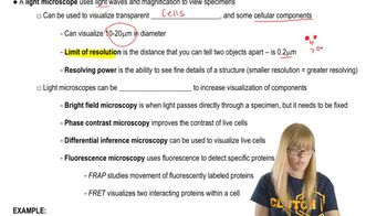
 03:43
03:43