 뒤로
뒤로Anatomy & Physiology: Blood and Heart Study Guide
 2:36
2:36
이 집합의 용어 (22)
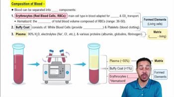
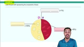
Blood consists of plasma (~55%) and formed elements (~45%).
Plasma transports nutrients, gases, hormones, and waste; contains proteins like albumin, globulins, and fibrinogen for osmotic balance, immunity, and clotting.
Formed elements include red blood cells, white blood cells, and platelets, comprising about 45% of whole blood.
Hematocrit measures the percentage of red blood cells in blood; typically higher in males (~47%) than females (~42%) due to hormonal differences.
Red blood cells are biconcave, lack nuclei, carry oxygen via hemoglobin, and have a lifespan of ~120 days.
White blood cells are granulocytes (neutrophils, eosinophils, basophils) and agranulocytes (lymphocytes, monocytes), involved in immune defense.
Platelets aid in blood clotting (hemostasis) and are formed from megakaryocytes in bone marrow.
Blood type depends on antigens (agglutinogens) on red cells and antibodies (agglutinins) in plasma, critical for safe transfusions.
Hemopoiesis is blood cell formation occurring mainly in the bone marrow.
The heart is located in the mediastinum, between the lungs, behind the sternum.
The heart has major arteries (aorta, pulmonary arteries) and veins (superior/inferior vena cava, pulmonary veins) connected externally.
Heart valves (tricuspid, pulmonary, mitral, aortic) ensure one-way blood flow through the heart chambers.
The right atrium receives deoxygenated blood; right ventricle pumps it to lungs; left atrium receives oxygenated blood; left ventricle pumps it to the body.
Pulmonary circulation moves blood between heart and lungs; systemic circulation moves blood between heart and body.
Coronary arteries supply the heart muscle; coronary veins drain deoxygenated blood from the myocardium.
Cardiac muscle has intercalated discs for electrical connectivity and striations for contraction.
Includes the sinoatrial (SA) node, atrioventricular (AV) node, bundle of His, bundle branches, and Purkinje fibers.
Impulse starts at SA node → AV node → bundle of His → bundle branches → Purkinje fibers, causing coordinated contraction.
ECG shows P wave (atrial depolarization), QRS complex (ventricular depolarization), and T wave (ventricular repolarization).
P wave represents atrial depolarization initiating atrial contraction.
QRS complex represents ventricular depolarization leading to ventricular contraction.
T wave represents ventricular repolarization, the recovery phase before the next heartbeat.