뒤로

Anatomy & Physiology: Digestive System and Metabolism

컨트롤 버튼이 '내비게이션' 모드로 변경되었습니다.
1/29
  • Anatomy of the mouth, lips, and palate

    The mouth includes lips, cheeks, tongue, hard palate, and soft palate. Lips protect the entrance, the hard palate forms the anterior roof, and the soft palate forms the posterior roof and closes the nasal passage during swallowing.
  • Functions of intrinsic and extrinsic tongue muscles

    Intrinsic muscles change tongue shape for speech and swallowing; extrinsic muscles alter tongue position for movement and food manipulation.
  • Three types of papillae on the tongue

    Filiform (texture), fungiform (taste buds), and circumvallate (large taste buds at the back).
  • Three pairs of extrinsic salivary glands

    Parotid, submandibular, and sublingual glands produce saliva to moisten food and begin digestion.
  • Functions and composition of saliva

    Saliva moistens food, begins starch digestion with amylase, cleanses mouth, and contains mucus, enzymes, electrolytes, and antibacterial agents.
  • Control of salivation

    Salivation is controlled by the autonomic nervous system; parasympathetic stimulation increases saliva, sympathetic reduces it.
  • Classification of teeth

    Incisors (cutting), canines (tearing), premolars and molars (grinding).
  • Structure of tooth crown and root

    Crown is covered by enamel; root is embedded in the jawbone and covered by cementum; both contain dentin and pulp.
  • Gomphosis joint

    A fibrous joint where the tooth root is anchored into the alveolar socket by the periodontal ligament.
  • Structure and functions of the pharynx

    A muscular tube connecting mouth to esophagus; functions in swallowing and passage of air to the larynx.
  • Soft palate position change during swallowing

    The soft palate elevates to close the nasopharynx, preventing food from entering the nasal cavity.
  • Muscle types in the esophagus

    Upper third: skeletal muscle; middle third: mixed skeletal and smooth muscle; lower third: smooth muscle.
  • Digestive process in the mouth

    Mechanical digestion by chewing and chemical digestion by salivary amylase breaking down starch.
  • Parts of the alimentary canal

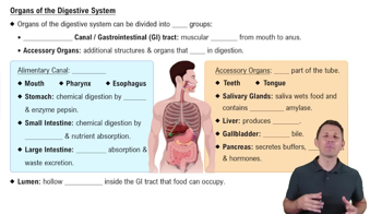
    Mouth, pharynx, esophagus, stomach, small intestine, large intestine, rectum, and anus.
  • Accessory digestive organs

    Teeth, tongue, salivary glands, liver, gallbladder, and pancreas.
  • Six essential digestive processes

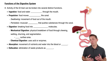
    Ingestion, propulsion, mechanical digestion, chemical digestion, absorption, and defecation; occurring throughout the alimentary canal.
  • Difference between peristalsis and segmentation

    Peristalsis moves food forward by wave-like contractions; segmentation mixes food by rhythmic contractions without net movement.
  • Intrinsic and extrinsic control of digestion

    Intrinsic control via enteric nervous system; extrinsic control via autonomic nervous system and hormones.
  • Short reflexes and long reflexes in digestion

    Short reflexes are local enteric responses; long reflexes involve CNS integration and autonomic output.
  • Mesentery and its formation

    A double layer of peritoneum that suspends intestines; formed as the gut tube rotates and folds during development.
  • Examples of peritoneal and retroperitoneal organs

    Peritoneal: stomach, liver, jejunum; retroperitoneal: pancreas, kidneys, ascending colon.
  • Hepatic portal circulation

    Blood from digestive organs flows through the hepatic portal vein to the liver for nutrient processing before entering systemic circulation.
  • Four tunics of the GI tract wall

    Mucosa, submucosa, muscularis externa, and serosa (or adventitia).
  • Enteric nervous system and its functions

    A network of neurons in the GI tract wall that controls motility, secretion, and blood flow independently.
  • Parts of the stomach

    Cardiac region, fundus, body, pyloric region, lesser curvature, and greater curvature.
  • Locations of greater and lesser omentum

    Greater omentum hangs from the greater curvature; lesser omentum connects the lesser curvature to the liver.
  • Blood supply of the stomach

    Branches of the celiac trunk including left and right gastric, left and right gastroepiploic, and short gastric arteries.
  • Muscular arrangements of the stomach wall

    Three layers: longitudinal, circular, and oblique muscles for mechanical digestion.
  • Major function of goblet cells

    Secrete mucus to protect and lubricate the mucosal lining.