Skip to main content
Anatomy & Physiology
나의 코스
배우기
시험 준비
AI 튜터
학습 가이드
교과서적 해결책
플래시카드
탐험해 보세요
앱을 사용해 보세요
나의 코스
배우기
시험 준비
AI 튜터
학습 가이드
교과서적 해결책
플래시카드
탐험해 보세요
앱을 사용해 보세요
뒤로
Anatomy & Physiology: Endocrine System and Blood
카드를 뒤집기 위해 탭할 수 있습니다.
Endocrine system function
카드를 뒤집기 위해 탭할 수 있습니다.
👆
Endocrine system function
Coordinates body cell activity via hormones transported in blood; controls reproduction, growth, metabolism, and defense mobilization.
진행 상황 추적
컨트롤 버튼이 '내비게이션' 모드로 변경되었습니다.
1/28
추천 영상
7:06
Components of the Endocrine System
13719
views
418
rank
6:20
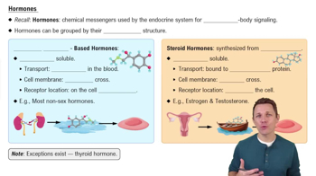
Hormones
10454
views
260
rank
3:33
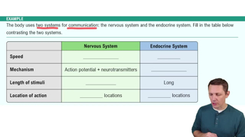
Introduction to the Endocrine System Example 1
14968
views
445
rank
1
comments
이 집합의 용어 (28)
하이드의 정의
Endocrine system function
Coordinates body cell activity via hormones transported in blood; controls reproduction, growth, metabolism, and defense mobilization.
Difference between endocrine and exocrine glands
Endocrine glands produce hormones and lack ducts; exocrine glands produce nonhormonal substances and have ducts.
Two main classes of hormones
Amino acid–based hormones (peptides, proteins) and steroids (synthesized from cholesterol).
Target cells in hormone action
Cells with specific receptors for a hormone; hormones alter their activity by changing membrane permeability, enzyme activity, secretion, or mitosis.
Three stimuli triggering hormone release
Humoral (blood ion/nutrient levels), neural (nerve fibers), and hormonal (hormones stimulating other glands).
Up-regulation vs. down-regulation of hormone receptors
Up-regulation: increase receptors due to low hormone levels; down-regulation: decrease receptors due to high hormone levels.
Half-life of hormones
Time for blood hormone level to decrease by half; varies from minutes to a week depending on hormone.
Permissiveness, synergism, and antagonism in hormone interaction
Permissiveness: one hormone needs another to act; synergism: hormones amplify effects; antagonism: hormones oppose each other.
Posterior pituitary hormones and origin
Oxytocin and ADH produced by hypothalamic neurons; stored and released by posterior pituitary.
Growth hormone (GH) effects
Direct metabolic effects (glucose sparing, fat breakdown) and indirect growth promotion via IGFs stimulating cell growth and protein synthesis.
Thyroid hormone forms and effects
T4 (thyroxine) and T3 (triiodothyronine); increase basal metabolic rate, regulate growth, development, and maintain blood pressure.
Parathyroid hormone (PTH) role
Increases blood Ca2+ by stimulating bone resorption, kidney reabsorption, and activating vitamin D for intestinal absorption.
Aldosterone function
Mineralocorticoid that stimulates Na+ reabsorption and K+ elimination by kidneys, increasing blood volume and pressure.
Renin-angiotensin-aldosterone mechanism
Low blood pressure triggers renin release, leading to angiotensin II formation, which stimulates aldosterone release.
Cortisol effects
Raises blood glucose via gluconeogenesis, promotes fat and protein metabolism, enhances vasoconstriction, and suppresses inflammation.
Adrenal medulla hormones and effects
Epinephrine and norepinephrine increase heart rate, blood glucose, and cause vasoconstriction; epinephrine stimulates metabolism more.
Pancreatic islet hormones
Alpha cells produce glucagon (raises blood glucose); beta cells produce insulin (lowers blood glucose).
Insulin actions
Enhances glucose uptake by cells, inhibits glycogen breakdown, and promotes glucose storage as glycogen or fat.
Erythrocyte structure
Biconcave, anucleate cells filled with hemoglobin for efficient oxygen transport.
Erythropoiesis regulation
Stimulated by erythropoietin (EPO) from kidneys in response to hypoxia; requires iron, vitamin B12, and folic acid.
Hemoglobin composition
Four polypeptide chains (two alpha, two beta) each with a heme group containing iron that binds oxygen.
Leukocyte categories
Granulocytes (neutrophils, eosinophils, basophils) and agranulocytes (lymphocytes, monocytes).
Neutrophil function
Most abundant WBC; phagocytizes bacteria and kills microbes via respiratory burst.
Platelet function
Cell fragments that form temporary plugs to seal vessel breaks and release chemicals for clotting.
Three steps of hemostasis
Vascular spasm, platelet plug formation, and coagulation (clotting).
Intrinsic vs. extrinsic coagulation pathways
Intrinsic triggered by blood factors; extrinsic triggered by tissue factor outside blood; both activate factor X.
Coagulation final steps
Prothrombin activator converts prothrombin to thrombin; thrombin converts fibrinogen to fibrin forming clot mesh.
Blood types ABO and Rh
ABO groups based on A and B antigens; Rh factor based on presence of D antigen; mismatches cause transfusion reactions.
 뒤로뒤로
뒤로뒤로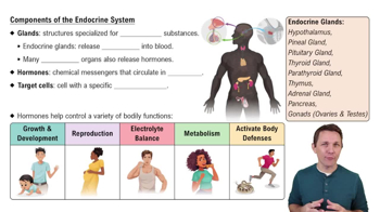
 7:06
7:06