 뒤로
뒤로Anatomy & Physiology Exam 3 Review: Nervous System
 07:53
07:53
이 집합의 용어 (25)
Efferent division carries motor impulses from the CNS to muscles and glands. Afferent division carries sensory impulses from receptors to the CNS.
Dendrites receive signals, axon transmits impulses away from the cell body, and myelin sheath insulates axons to speed up impulse conduction.
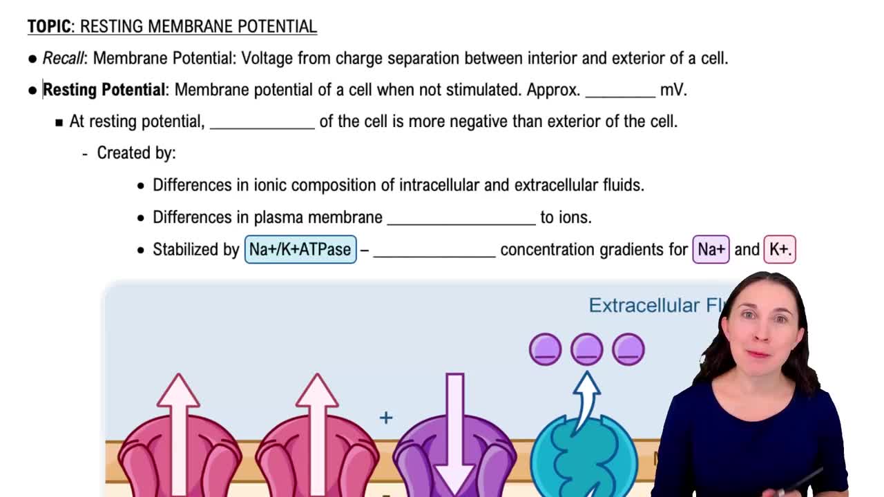
The inside of the cell is more negative than the outside at resting membrane potential.
Ependymal produce cerebrospinal fluid, oligodendrocytes myelinate CNS axons, and microglia act as CNS immune cells.
Graded potentials vary in size and occur in dendrites; action potentials are all-or-none signals that propagate along axons.
Includes depolarization, repolarization, and hyperpolarization phases, reflecting changes in membrane potential.
CSF circulates through the ventricles, central canal of spinal cord, and subarachnoid space around brain and spinal cord.
Choroid plexuses produce cerebrospinal fluid within the brain ventricles.
The meninges consist of pia mater (innermost), arachnoid mater (middle), and dura mater (outermost).
Gray matter contains neuron cell bodies; white matter contains myelinated axons.
Specific CNS regions process sensory input, including the dorsal parts of the spinal cord and sensory cortex.
Dorsal parts contain sensory neurons; central parts include gray matter and interneurons.
The spinal cord ends at the L2 vertebra.
The end of the spinal cord is called the conus medullaris.
Gray matter processes information; white matter transmits signals up and down the spinal cord.
The PNS is made up of cranial nerves, spinal nerves, and associated ganglia.
Dermatomes are skin areas supplied by sensory nerves; myotomes are muscle groups controlled by motor nerves.
The vagus nerve primarily decreases heart rate and promotes digestion (parasympathetic effects).
Somatic controls voluntary skeletal muscles; autonomic controls involuntary functions of organs and glands.
Includes autonomic (involuntary organ control), somatic (skeletal muscle), flexor (withdrawal), and plantar reflexes.
Muscle spindles detect changes in muscle length and trigger stretch reflexes.
Golgi tendon organs sense muscle tension to prevent muscle damage from excessive force.
The ANS has two divisions: sympathetic (fight or flight) and parasympathetic (rest and digest).
Most organs receive input from both sympathetic and parasympathetic nerves for balanced control.
Parasympathetic nerves leave the spinal cord from the craniosacral regions.