 뒤로
뒤로Anatomy & Physiology II: Cardiovascular System Study Guide
 2:36
2:36
이 집합의 용어 (26)
Hematology is the study of blood.
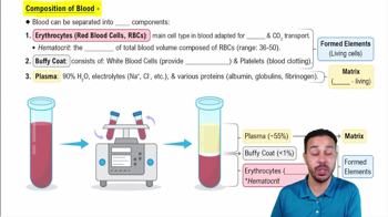
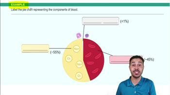
Blood is made of plasma (liquid portion) and formed elements (cells and cell fragments).
Plasma makes up approximately 46–63% of whole blood.
The three types are red blood cells, white blood cells, and platelets.
To transport oxygen from the lungs to tissues and some carbon dioxide from tissues to lungs.
Red blood cells are biconcave discs, lack a nucleus, mitochondria, and ribosomes, and appear pink with pale centers on a smear.
Approximately 120 days.
White blood cells are called leukocytes and are involved in body defense against pathogens and abnormal cells.
Neutrophils, eosinophils, basophils, monocytes, and lymphocytes.
Neutrophils (50–70%) perform phagocytosis to destroy bacteria and debris.
Never Let Monkeys Eat Bananas (Neutrophils, Lymphocytes, Monocytes, Eosinophils, Basophils).
Platelets, or thrombocytes, are involved in blood clotting.
Right atrium, right ventricle, left atrium, and left ventricle.
The right side pumps to the lungs; the left side pumps to the body.
Atria are receiving chambers; ventricles are pumping chambers.
Venae cavae → right atrium → tricuspid valve → right ventricle → pulmonary semilunar valve → pulmonary trunk → pulmonary arteries → lungs → pulmonary veins → left atrium → mitral valve → left ventricle → aortic semilunar valve → aorta → body.
Pulmonary arteries carry oxygen-poor blood; pulmonary veins carry oxygen-rich blood, opposite of usual arteries and veins.
Tricuspid (right atrium to right ventricle), mitral/bicuspid (left atrium to left ventricle), pulmonary semilunar (right ventricle to pulmonary trunk), aortic semilunar (left ventricle to aorta).
They prevent AV valves from flipping backward during ventricular contraction.
The SA node is the heart's natural pacemaker located in the right atrium; it initiates the electrical impulse for heartbeat.
SA node → AV node → AV bundle (Bundle of His) → right and left bundle branches → Purkinje fibers.
The P wave represents atrial depolarization.
The QRS complex represents ventricular depolarization.
The T wave represents ventricular repolarization.
The PR interval is the time from the start of atrial depolarization to the start of ventricular depolarization, including AV node delay.
The RR interval measures time between heartbeats and is used to calculate heart rate; shorter RR means faster rate, longer means slower.