Transport of gases and nutrients, regulation of pH and temperature, and protection against infection and blood loss.
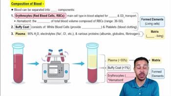
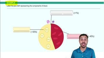
Major component of blood
Water makes up the majority of blood volume.
Hematocrit definition
The percentage of red blood cells (RBCs) relative to the total blood volume.
Buffy coat layer
The layer in centrifuged blood containing white blood cells and platelets.
Agranulocytes vs granulocytes
Agranulocytes have no granules (lymphocytes, monocytes); granulocytes have granules (neutrophils, eosinophils, basophils).
Most abundant white blood cell
Neutrophil is the most common type of white blood cell.
When does hemopoiesis occur?
During low oxygen (hypoxia) or when there are low red blood cells.
Myeloid vs lymphoid stem cells
Myeloid stem cells produce granulocytes; lymphoid stem cells produce agranulocytes (T and B lymphocytes).
Erythropoietin (EPO) source and function
Produced by the kidney, EPO stimulates bone marrow to produce more red blood cells.
Where is oxygen held in RBCs?
Oxygen binds to the heme group of hemoglobin, which contains iron.
Characteristics of red blood cells
Biconcave discs, filled with hemoglobin, lack organelles, contain spectrin, and transport gases.
Characteristics of leukocytes
True cells with organelles; some have granules; function in protection.
ABO blood group antigens and antibodies
Antigens on RBC surface; antibodies in plasma. Group A has A antigen and anti-B antibodies; B has B antigen and anti-A antibodies; AB has both antigens and no antibodies; O has no antigens and both anti-A and anti-B antibodies.
Universal donor and receiver blood types
O is universal donor; AB is universal receiver.
Erythroblastosis fetalis cause
Rh-negative mother sensitized to Rh-positive fetus blood; antibodies attack second Rh-positive fetus's RBCs causing hemolysis.
How do platelets adhere to vascular walls?
Platelets adhere when collagen is exposed at injury sites.
Layers of the pericardium
From outside in: fibrous pericardium, parietal serous pericardium, visceral serous pericardium with pericardial fluid between parietal and visceral layers.
Layers of the heart wall
Epicardium (same as visceral serous pericardium), myocardium, and endocardium (lines chambers).
Characteristics of cardiac muscle tissue
Striated, branched, short cells with one central nucleus and intercalated discs containing gap junctions.
Function of chordae tendineae
Attach papillary muscles to AV valves to prevent valve prolapse during ventricular contraction.
Why is the left ventricle wall thicker than the right?
Left ventricle pumps blood to the entire body requiring higher pressure; right ventricle pumps only to lungs at lower pressure.
Fossa ovalis significance
Remnant of fetal foramen ovale; an indentation in the interatrial septum after birth.
When are AV valves open and closed?
Open during ventricular diastole (relaxation) to allow filling; closed during ventricular systole (contraction) to prevent backflow.
Location of semilunar valves
Aortic valve at the aorta; pulmonary valve at the pulmonary trunk.
Heart valve auscultation points
Tricuspid: 5th intercostal space right sternum; Mitral: 5th intercostal space left sternum; Aortic: 2nd intercostal space right sternum; Pulmonary: 2nd intercostal space left sternum.
Conduction system of the heart
SA node (pacemaker), AV node (delay), Bundle of His, bundle branches, Purkinje fibers; ventricles contract when impulse reaches Purkinje fibers.
Why is impulse delayed at AV node?
To ensure atria finish contracting before ventricles contract.
ECG wave meanings
P wave: atrial depolarization; QRS complex: ventricular depolarization; T wave: ventricular repolarization.
Cardiac output (CO) formula
CO = \(SV \times HR\) (Stroke Volume times Heart Rate).
Stroke volume (SV) formula
SV = EDV - ESV (End Diastolic Volume minus End Systolic Volume).
Frank-Starling law of the heart
The more the ventricles are stretched during filling (preload), the greater the force of contraction and blood ejected.
Differences between arteries and veins
Arteries have thicker walls, more smooth muscle and elastin, smaller lumen, and no valves; veins have thinner walls, larger lumen, less muscle, and valves to prevent backflow.
Tunics of blood vessels
From inside out: Tunica interna (endothelium), Tunica media (smooth muscle), Tunica externa (connective tissue).
Characteristics of capillaries
One cell thick endothelium (tunica interna), permeable for diffusion and filtration.
Which veins carry oxygenated blood?
Pulmonary veins carry oxygenated blood from lungs to heart.
Why is blood flow velocity slowest in capillaries?
Because capillaries have the greatest total cross-sectional area.
Organ with constant blood flow
The brain requires constant blood flow and is intolerant to ischemia.
 뒤로
뒤로
 2:36
2:36