뒤로

Anatomy & Physiology Midterm Practical Key Concepts

컨트롤 버튼이 '내비게이션' 모드로 변경되었습니다.
1/28
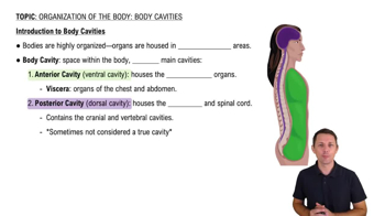
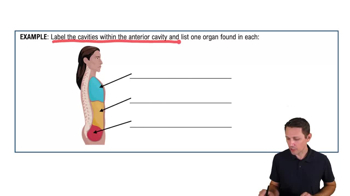
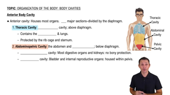
  • Name the major body cavities in the torso.

    Thoracic, abdominopelvic, cranial, spinal, pleural, pericardial, and mediastinal cavities.
  • What muscle separates the thoracic and abdominopelvic cavities?

    Diaphragm muscle.
  • How are the 4 abdominal quadrants defined?

    Using a tape grid on chest plates dividing the abdomen into right upper, left upper, right lower, and left lower quadrants.
  • What are the 9 abdominal regions used for anatomical reference?

    Divisions using a tape grid: right and left hypochondriac, epigastric, right and left lumbar, umbilical, right and left iliac, and hypogastric regions.
  • List the 4 primary classes of tissue to recognize under the microscope.

    Epithelial, connective, muscle, and nervous tissues.
  • Name three types of cartilage tissue to identify.

    Fibrous, elastic, and hyaline cartilage.
  • What are the two types of bone tissue to distinguish microscopically?

    Compact bone and spongy (cancellous) bone.
  • How can you differentiate white blood cells from red blood cells in a blood smear?

    White blood cells have nuclei and are larger; red blood cells lack nuclei and are smaller, biconcave discs.
  • What are the differences between DICT and DRCT connective tissues?

    DICT (Dense Irregular Connective Tissue) has fibers in multiple directions; DRCT (Dense Regular Connective Tissue) has fibers aligned parallel.
  • Name the layers of the epidermis to identify on skin models.

    Stratum basale, stratum spinosum, stratum granulosum, stratum lucidum (thick skin only), and stratum corneum.
  • What glands are found in the skin and their functions?

    Sebaceous glands (oil), sudoriferous glands (sweat), and apocrine glands (axillary sweat).
  • Identify the parts of a hair follicle on skin models.

    Hair bulb, root, and shaft.
  • What are the key features of an osteon in bone tissue?

    Concentric lamellae, central canal, osteocytes in lacunae, and canaliculi.
  • Differentiate compact bone from spongy bone structurally.

    Compact bone is dense with osteons; spongy bone has trabeculae and marrow spaces.

  • Name the main parts of a long bone to identify.

    Epiphyses (proximal and distal), diaphysis, articular surfaces, marrow/medullary cavity, and spongy bone.
  • How to distinguish cervical, thoracic, and lumbar vertebrae?

    Cervical vertebrae have transverse foramina; thoracic have facets for ribs; lumbar are larger with thick bodies.
  • Name the four rotator cuff muscles.

    Supraspinatus, infraspinatus, subscapularis, and teres minor.
  • List the muscles of the quadriceps group.

    Vastus lateralis, vastus intermedius, vastus medialis, and rectus femoris.
  • What are the main external features of the heart to identify?

    Right and left atria, right and left ventricles, pulmonary arteries and veins, aorta, aortic arch, superior and inferior vena cava, and pericardium.
  • Name the interior heart structures important for valve function.

    Chordae tendineae, papillary muscles, right and left cuspid valves, right and left semilunar valves, trabeculae carneae.
  • What are the three layers of blood vessel walls?

    Tunica intima (endothelium), tunica media (smooth muscle), and tunica externa (adventitia).
  • How to distinguish arteries from veins microscopically?

    Arteries have thicker tunica media and smaller lumen; veins have thinner walls and larger lumen.
  • Identify the sutures of the skull.

    Sagittal, lambdoid, coronal, and squamous sutures.
  • Name the cranial bones.

    Frontal, parietal, temporal, occipital, ethmoid, and sphenoid bones.
  • Name the facial bones listed in the study materials.

    Maxilla, zygomatic, nasal, lacrimal, palatine, vomer, and inferior nasal conchae.
  • What are the key landmarks of the sphenoid bone?

    Sella turcica, greater wings, and lesser wings.
  • What are the key landmarks of the temporal bone?

    External acoustic meatus, mastoid process, styloid process, petrous part, mandibular fossa.
  • How to distinguish male from female pelvis features?

    Male pelvis is narrower with a smaller pelvic inlet and sharper pubic angle; female pelvis is wider with a larger inlet and broader pubic angle.