Skip to main content
Anatomy & Physiology
나의 코스
배우기
시험 준비
AI 튜터
학습 가이드
교과서적 해결책
플래시카드
탐험해 보세요
앱을 사용해 보세요
나의 코스
배우기
시험 준비
AI 튜터
학습 가이드
교과서적 해결책
플래시카드
탐험해 보세요
앱을 사용해 보세요
뒤로
Anatomy & Physiology: Skeletal and Nervous System Essentials
카드를 뒤집기 위해 탭할 수 있습니다.
Frontal Bone
카드를 뒤집기 위해 탭할 수 있습니다.
👆
Frontal Bone
The bone forming the forehead and the upper part of the eye sockets.
진행 상황 추적
컨트롤 버튼이 '내비게이션' 모드로 변경되었습니다.
1/29
추천 영상
2:12
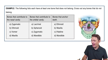
The Skull Example 1
10235
views
237
rank
4:42
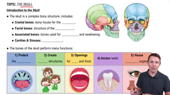
Introduction to the Skull
18549
views
422
rank
3:40
The Skull Example 3
7418
views
144
rank
이 집합의 용어 (29)
하이드의 정의
Frontal Bone
The bone forming the forehead and the upper part of the eye sockets.
Sagittal Suture
The fibrous joint connecting the two parietal bones of the skull.
Mastoid Process
A bony projection of the temporal bone located behind the ear.
Foramen Magnum
The large opening at the base of the skull through which the spinal cord passes.
C1 (Atlas)
The first cervical vertebra that supports the skull and allows nodding motion.
Dens (C2 only)
A tooth-like projection on the axis (C2) that allows head rotation.
True Ribs
The first seven pairs of ribs that attach directly to the sternum via costal cartilage.
Xiphoid Process
The small, cartilaginous lower tip of the sternum.
Acetabulum
The deep socket in the hip bone that receives the head of the femur.
Coracoid Process
A hook-like bony projection on the scapula serving as an attachment point for muscles.
Greater Tubercle of Humerus
A large lateral projection on the humerus for muscle attachment.
Olecranon Fossa
A depression on the posterior humerus that accommodates the olecranon of the ulna.
Scaphoid
A carpal bone located near the base of the thumb in the wrist.
Fovea Capitis
A small pit on the head of the femur for ligament attachment.
Medial Cuneiform
A tarsal bone located on the medial side of the foot.
Orbicularis Oculi
A muscle that encircles the eye and allows blinking and squinting.
Deltoid Muscle
The thick, triangular shoulder muscle responsible for arm abduction.
Rectus Femoris
A quadriceps muscle that extends the knee and flexes the hip.
External Obliques
Muscles on the lateral sides of the abdomen that help rotate the trunk.
Corpus Callosum
A thick band of nerve fibers connecting the two cerebral hemispheres.
Thalamus
A brain structure that relays sensory and motor signals to the cerebral cortex.
Pons
Part of the brainstem involved in motor control and sensory analysis.
Arbor Vitae
The tree-like white matter structure inside the cerebellum.
Optic Chiasma
The X-shaped structure where optic nerves partially cross.
Dorsal Root Ganglion
A cluster of sensory neuron cell bodies located on the dorsal root of a spinal nerve.
Ventral Horn
The front column of gray matter in the spinal cord containing motor neurons.
External Acoustic Meatus
The ear canal leading to the eardrum.
Jugular Notch
A visible dip at the superior border of the manubrium of the sternum.
Ischium
The lower, posterior part of the hip bone that supports body weight when sitting.
 뒤로뒤로
뒤로뒤로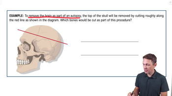
 2:12
2:12