 뒤로
뒤로Anatomy & Physiology: Special Senses
 5:05
5:05
이 집합의 용어 (28)
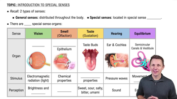
The five special senses are olfaction (smell), gustation (taste), vision, hearing, and vestibular sensation.
Special sense receptors are usually specialized cells rather than neurons and transmit stimuli to the CNS via cranial nerves only.
Transduction is the process where a physical or chemical stimulus is converted into an action potential.
The olfactory epithelium is in the roof of the nasal cavity and contains olfactory neurons whose axons form the olfactory nerve (CN I).
Receptors on the cilia of olfactory neurons bind to odorant molecules.
In the primary olfactory cortex, with signals also sent to the limbic system for emotional and visceral responses.
Taste buds are located on the vallate, fungiform, and foliate papillae of the tongue.
There are five: sweet, salty, sour, bitter, and umami (savory).
Taste stimuli are transmitted via neurons of the facial (VII), glossopharyngeal (IX), and vagus (X) nerves.
In the parietal lobe for conscious awareness and the limbic system for emotional and visceral responses.
The fibrous layer (sclera and cornea), vascular layer (choroid, ciliary body, iris), and neural layer (retina).
The lens alters the refraction of light to focus on near and far objects by changing shape.
The posterior cavity contains vitreous humor, and the anterior cavity contains aqueous humor.
Rods detect light intensity (low light), and cones detect color in three types sensitive to different wavelengths.
Accommodation is the lens's ability to become rounder to focus light on the retina for near vision.
They hyperpolarize, removing inhibition on bipolar cells, which then stimulate retinal ganglion cells to send signals via the optic nerve.
They cross at the optic chiasma, allowing visual information from the nasal retina to be processed by the opposite brain hemisphere.
The outer ear, middle ear, and inner ear.
The tympanic membrane, pharyngotympanic tube, and three auditory ossicles: malleus, incus, and stapes.
Endolymph is inside the membranous labyrinth; perilymph surrounds it between the membranous labyrinth and bone.
The cochlea contains structures for hearing, including the cochlear duct and scala vestibuli and tympani, where sound vibrations are transduced.
Sound waves vibrate the tympanic membrane and ossicles, moving the basilar membrane and hair cells in the cochlea, which release neurotransmitters to cochlear nerve neurons.
In the primary auditory cortex of the temporal lobe, with further processing in temporal, parietal lobes, and limbic system.
They detect head tilting and linear acceleration by displacement of the otolithic membrane bending hair cells.
Rotation moves endolymph against the cupula, bending hair cells' stereocilia and kinocilium, causing depolarization or hyperpolarization.
In the spinal cord, cerebellum, inferior parietal lobe, and cranial nerve nuclei for eye movement coordination.
Stimuli travel from the brainstem to the thalamus and then to the primary sensory cortex.
The frontal lobe integrates sensory information into a coherent picture, and the limbic system provides emotional and behavioral responses.