 뒤로
뒤로Anatomy & Physiology: Special Senses - Eye Anatomy and Physiology
 5:05
5:05
이 집합의 용어 (28)
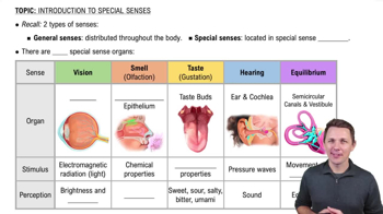
Smell, taste, sight, hearing, and equilibrium are the five special senses.
About 70 percent of all sensory receptors are located in the eyes.
Accessory structures include the extrinsic eye muscles, eyelids, conjunctiva, and lacrimal apparatus.
Tarsal glands produce an oily secretion that lubricates the eye.
The conjunctiva is a membrane lining the eyelids and eyeball that secretes mucus to lubricate and keep the eye moist.
The lacrimal apparatus includes the lacrimal gland and ducts. Tears flow from the gland across the eye into lacrimal canaliculi, then lacrimal sac, nasolacrimal duct, and finally the nasal cavity.
Tears contain dilute salt solution, mucus, antibodies, and lysozyme, which cleanse, protect, moisten, and lubricate the eye.
There are six extrinsic eye muscles that attach to the eye's outer surface and produce gross eye movements.
The eyeball wall has three layers: fibrous (outer), vascular (middle), and sensory (inner).
The fibrous layer consists of the sclera (white connective tissue) and the cornea (transparent anterior portion).
The choroid is a blood-rich layer that nourishes the eye and contains pigment to prevent light scattering.
The ciliary body (attached to the lens) and the iris (regulates light entry) are derived from the choroid.
The retina has an outer pigmented layer that absorbs light and an inner neural layer containing photoreceptors (rods and cones).
Rods detect dim light and peripheral vision, providing gray tone perception.
Cones allow detailed color vision and are densest in the fovea centralis, the area of sharpest vision.
The optic disc is the blind spot where the optic nerve exits the eye; no photoreceptors are present here.
The anterior segment contains aqueous humor, and the posterior segment contains vitreous humor.
Aqueous humor maintains intraocular pressure, nourishes the lens and cornea, and is reabsorbed via the scleral venous sinus.
Vitreous humor is a gel-like substance that prevents eye collapse and helps maintain intraocular pressure.
Light is refracted by the cornea, aqueous humor, lens, and vitreous humor to focus on the retina.
Accommodation is the lens changing shape to focus on objects closer than 20 feet.
The image on the retina is a real image that is reversed left to right, upside down, and smaller than the object.
Visual signals travel from the retina → optic nerve → optic chiasma → optic tract → thalamus → optic radiation → visual cortex.
Binocular vision results from overlapping visual fields of both eyes and provides depth perception.
Myopia (nearsightedness) results from an eyeball that is too long, causing distant objects to focus in front of the retina and appear blurry.
Hyperopia (farsightedness) results from an eyeball that is too short or a lazy lens, causing near objects to focus behind the retina and appear blurry.
Astigmatism causes blurry images due to unequal curvatures of the cornea or lens, focusing light as lines instead of points.
Convergence (eyes move medially for close focus), photopupillary reflex (pupils constrict in bright light), and accommodation pupillary reflex (pupils constrict when viewing close objects).