 뒤로
뒤로Anatomy & Physiology: The Spinal Cord and CNS Pathways
 0:50
0:50
이 집합의 용어 (20)
The spinal cord is a long, tubular organ encased within the vertebral cavity, beginning at the foramen magnum and ending between the first and second lumbar vertebrae.
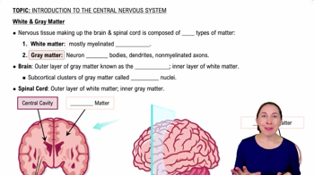
The central canal is filled with cerebrospinal fluid (CSF) and is continuous with the brain's ventricles, providing cushioning and nutrient transport.
White matter contains myelinated axons and is superficial in the spinal cord; gray matter contains neuron cell bodies, dendrites, and unmyelinated axons and is internal.
It acts as a relay station for communication between the brain and body, and as a processing station for spinal reflexes independent of the brain.
The meninges are the dura mater, arachnoid mater, and pia mater. The spinal dura mater lacks a periosteal layer, consisting only of a meningeal layer.
Denticulate ligaments extend from the pia mater through the arachnoid to the dura mater, anchoring the spinal cord within the vertebral cavity.
Epidural space (between dura and vertebrae, contains fat and veins), subdural space (potential space between dura and arachnoid), and subarachnoid space (between arachnoid and pia, filled with CSF).
Injection of anesthetic into the epidural space to numb nerves below the injection site, commonly used during childbirth and surgeries.
Between the fourth and fifth lumbar vertebrae to withdraw CSF safely without injuring the spinal cord, used to diagnose infections or diseases.
Bulges in the spinal cord where nerve roots for the upper and lower limbs emerge, reflecting increased neural input/output.
The conus medullaris is the tapered end of the spinal cord; the filum terminale is a thin extension of pia mater anchoring the cord to the coccyx.
Anterior horn (somatic motor neurons), posterior horn (sensory neurons), and lateral horn (autonomic motor neurons, thoracic to lumbar levels).
Contains cell bodies of sensory neurons whose axons enter the posterior horn of the spinal cord.
Regions of white matter on each side of the spinal cord: posterior, lateral, and anterior funiculi, containing ascending and descending tracts.
Posterior columns (fasciculus gracilis and cuneatus) carry touch and proprioception; spinocerebellar tracts carry proprioceptive info to cerebellum; anterolateral system carries pain and temperature.
The largest descending motor tract controlling skeletal muscles below the head and neck, originating in the cerebral cortex and decussating in the medulla.
First-order neurons detect stimuli and send to spinal cord; second-order neurons relay to thalamus; third-order neurons deliver impulses to cerebral cortex for interpretation.
Upper motor neurons (cerebral cortex), interneurons (brainstem/spinal cord), and lower motor neurons (anterior horn to muscles).
Upper motor neurons descend from motor cortex, decussate at medullary pyramids, travel in lateral funiculi, and synapse on lower motor neurons in anterior horn.
Control muscles of the head and neck via cranial nerve lower motor neurons; fibers do not decussate but synapse with brainstem interneurons.