 뒤로
뒤로Anatomy & Physiology: The Urinary System

 0:54
0:54
이 집합의 용어 (30)
Dispose of waste products like nitrogenous wastes, toxins, drugs, and excess ions; regulate blood pressure via renin; stimulate red blood cell production via erythropoietin; and activate vitamin D.
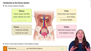
Kidneys, ureters, urinary bladder, and urethra make up the urinary system.
Located retroperitoneally from vertebrae T12 to L3; the right kidney is lower due to the liver's position.
Fibrous capsule, perirenal fat capsule, and renal fascia protect and anchor the kidney.
Renal cortex (outer), renal medulla (with pyramids and columns), and renal pelvis (funnel-shaped medial region).
Renal artery → segmental arteries → interlobar arteries → arcuate arteries → cortical radiate arteries.
Cortical radiate veins → arcuate veins → interlobar veins → renal vein → inferior vena cava; no segmental veins.
Each nephron has a renal corpuscle (glomerulus and Bowman's capsule) and a renal tubule (PCT, loop of Henle, DCT).
Cortical nephrons are mostly in the cortex; juxtamedullary nephrons have loops that dip deep into the medulla.
Specialized for filtration; high pressure forces water and solutes out of blood into Bowman's capsule.
Low-pressure capillaries adapted for reabsorption of water and solutes from renal tubules back into blood.
Glomerular filtration, tubular reabsorption, and tubular secretion.
A nonselective passive process where water and small solutes pass through the glomerular membrane into Bowman's capsule, forming filtrate.
Most reabsorption of water, glucose, amino acids, and ions occurs in the proximal convoluted tubule.
Removes substances like hydrogen and potassium ions, creatinine, and drugs from blood into renal tubules to maintain acid-base balance and eliminate wastes.
Urea (protein breakdown), uric acid (nucleic acid metabolism), and creatinine (muscle metabolism).
Clear, pale to deep yellow due to urochrome pigment, sterile at formation, slightly aromatic, slightly acidic (pH ~6), specific gravity 1.001–1.035.
Sodium, potassium, urea, uric acid, creatinine, ammonia, and bicarbonate ions.
Glucose, blood proteins, red blood cells, hemoglobin, white blood cells, bile.
Slender tubes that transport urine from kidneys to urinary bladder using peristalsis and gravity.
Muscular sac with detrusor muscle walls and transitional epithelium mucosa; stores urine temporarily; trigone region marks ureter and urethra openings.
Two sphincters: internal urethral sphincter (involuntary, smooth muscle) and external urethral sphincter (voluntary, skeletal muscle).
Bladder stretch receptors send impulses to spinal cord; pelvic nerves cause bladder contraction; urine passes internal sphincter; external sphincter is voluntarily controlled.
Excrete nitrogen wastes, maintain water balance, maintain electrolyte balance, and ensure proper blood pH.
Two-thirds intracellular fluid (ICF), one-third extracellular fluid (ECF) including plasma, interstitial fluid, lymph, and transcellular fluid.
Osmoreceptors in hypothalamus detect plasma solute concentration; dry mouth also stimulates thirst center to promote water intake.
Antidiuretic hormone (ADH) increases water reabsorption in collecting ducts to prevent water loss.
Stimulates sodium reabsorption and potassium secretion in kidneys; water follows sodium, increasing blood volume and pressure.
Low blood pressure triggers renin release from juxtaglomerular cells; renin produces angiotensin II causing vasoconstriction and aldosterone release.
Normal pH 7.35–7.45; alkalosis pH > 7.45; acidosis pH < 7.35; kidneys help maintain this balance.