 뒤로
뒤로Anatomy & Physiology: Tissue Types and Integumentary System
 03:32
03:32
이 집합의 용어 (22)
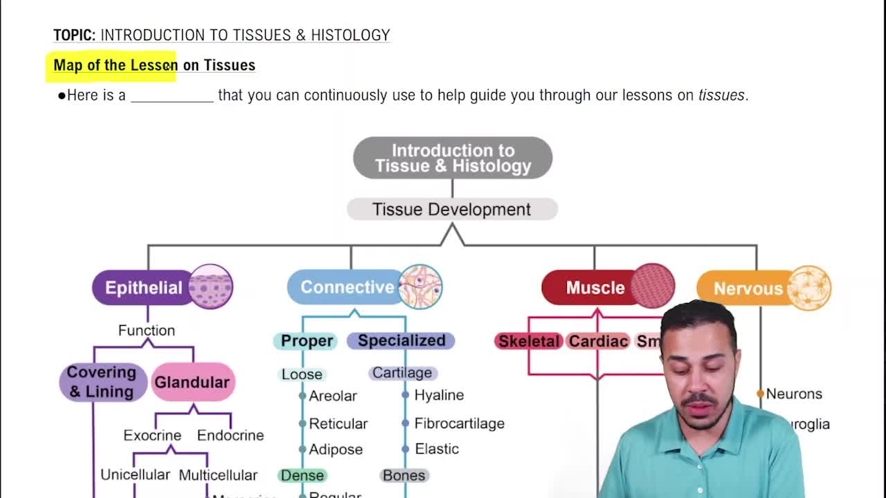
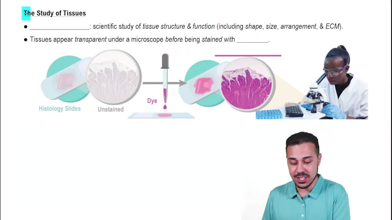
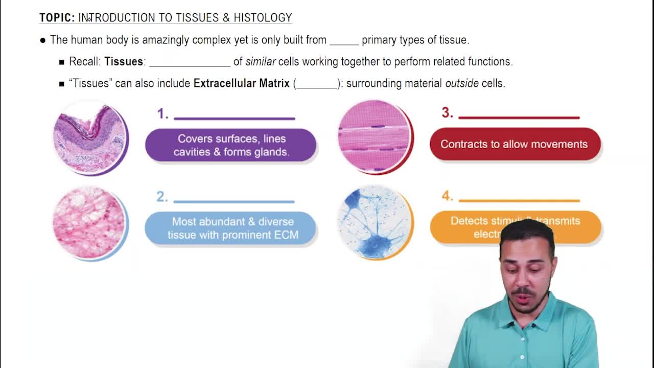
A group of similar cells organised to perform a specific function. Cells interact with extracellular matrix and signalling systems. Organs are formed by combinations of different tissues.
1. Epithelial tissue: covering, lining, secretion
2. Connective tissue: support, binding, transport
3. Nervous tissue: communication and control
4. Muscle tissue: contraction and movement
Sheets of closely packed cells, little extracellular matrix, apical-basal polarity, avascular, rapid regeneration.
Lines internal and external surfaces, forms protective layers, forms glands, and performs secretion.
Simple epithelium: one cell layer for exchange and absorption.
Stratified epithelium: multiple layers for protection.
Shapes: squamous, cuboidal, columnar.
Specialised: ciliated and glandular epithelium.
Cells separated by abundant extracellular matrix containing fibres (collagen, elastic, reticular) and ground substance. Usually well vascularised.
Protection, support, binding organs together, transport, and immunity.
Resident cells: fibroblasts, adipocytes, macrophages, mast cells, adult stem cells.
Other cells: chondrocytes (cartilage), osteocytes (bone), leukocytes (blood).
Loose connective tissue: fewer cells and fibres, more ground substance.
Dense connective tissue: more cells and fibres, less ground substance (e.g., tendons).
Adipose tissue (fat), compact bone (rigid), fibrocartilage (rigid yet flexible), blood (fluid connective tissue).
Skeletal muscle: voluntary, striated, multinucleated, attached to bones.
Smooth muscle: involuntary, non-striated, spindle-shaped, found in internal organs.
Cardiac muscle: involuntary, striated, single central nucleus, found in heart walls.
Specialised for rapid communication, excitable, capable of sending and receiving electrical signals. Main cells are neurons and glial cells.
Skin, hair, nails, sweat glands, sebaceous glands, cutaneous blood vessels, sensory receptors.
Protection, thermoregulation, sensation, vitamin D synthesis, and excretion.
Epidermis: keratinised stratified squamous epithelium, avascular.
Dermis: dense connective tissue with blood vessels, nerves, glands.
Hypodermis: subcutaneous tissue with adipose and loose connective tissue.
Keratinocytes, melanocytes, Langerhans cells, Merkel cells.
Mechanoreceptors (touch, pressure), thermoreceptors (temperature), nociceptors (pain), proprioceptors (body position), chemoreceptors (chemical stimuli).
Merkel discs: slow-adapting, sustained touch.
Meissner corpuscles: fast-adapting, texture changes.
Ruffini endings: slow-adapting, skin stretch.
Pacinian corpuscles: fast-adapting, deep pressure.
Vasodilation increases blood flow and sweating for heat loss.
Vasoconstriction reduces blood flow and sweating to conserve heat.
Piloerection causes hairs to stand; pilorelaxation causes hairs to flatten.
Melanin (eumelanin: brown/black, pheomelanin: red/yellow), carotene (dietary), haemoglobin (red blood cells).
Erythema: redness from inflammation or infection.
Pallor: paleness from anemia or poor circulation.
Jaundice: yellowing from bilirubin buildup.
Cyanosis: bluish from low oxygen.
Ecchymosis: bruising from bleeding disorders.
1. Haemostasis: clot formation.
2. Inflammation: white blood cells clear debris.
3. Proliferation: collagen scaffolding and new blood vessels.
4. Maturation: collagen reorganised, scar fades.