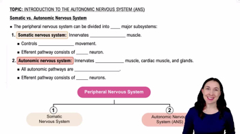
The autonomic division controls involuntary body functions by regulating smooth muscle, cardiac muscle, and glands to maintain homeostasis.
Branches of the autonomic division
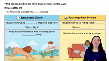
The autonomic division has two main branches: the sympathetic division, which prepares the body for 'fight or flight,' and the parasympathetic division, which promotes 'rest and digest' activities.
Anatomy of the sympathetic branch
Sympathetic neurons originate in the thoracolumbar spinal cord and have short preganglionic and long postganglionic fibers.
Anatomy of the parasympathetic branch
Parasympathetic neurons originate in the brainstem and sacral spinal cord with long preganglionic and short postganglionic fibers.
Chemical communication in the parasympathetic branch
Both preganglionic and postganglionic parasympathetic neurons release acetylcholine.
Synthesis of autonomic neurotransmitters
Acetylcholine is synthesized from choline and acetyl-CoA; norepinephrine is synthesized from dopamine in sympathetic neurons.
Breakdown of acetylcholine
Acetylcholine is broken down by the enzyme acetylcholinesterase in the synaptic cleft.
Breakdown of norepinephrine
Norepinephrine is removed by reuptake into the neuron and degraded by monoamine oxidase (MAO) and catechol-O-methyltransferase (COMT).
Structure of the adrenal medulla
The adrenal medulla is a modified sympathetic ganglion that secretes catecholamines directly into the bloodstream.
Secretions of the adrenal medulla
It secretes mainly epinephrine and some norepinephrine to enhance sympathetic responses.
Structure of the neuromuscular junction
The neuromuscular junction consists of a motor neuron terminal, synaptic cleft, and motor end plate on the muscle fiber.
Neurotransmitter at the neuromuscular junction
Acetylcholine is released from the motor neuron to stimulate muscle contraction.
Receptors at the neuromuscular junction
Nicotinic acetylcholine receptors on the muscle fiber membrane bind acetylcholine to initiate contraction.
Comparison of somatic motor and autonomic divisions - anatomy
Somatic motor neurons have a single long axon to skeletal muscle; autonomic neurons have two neurons (preganglionic and postganglionic) to target tissues.
Comparison of neurotransmitters in somatic motor vs autonomic divisions
Somatic motor neurons release acetylcholine at neuromuscular junctions; autonomic neurons release acetylcholine or norepinephrine depending on branch.
Receptors in somatic motor division
Somatic motor neurons use nicotinic acetylcholine receptors on skeletal muscle fibers.
Receptors in sympathetic division
Sympathetic postganglionic neurons release norepinephrine that binds to adrenergic receptors; preganglionic neurons release acetylcholine to nicotinic receptors.
Receptors in parasympathetic division
Parasympathetic postganglionic neurons release acetylcholine that binds to muscarinic receptors; preganglionic neurons release acetylcholine to nicotinic receptors.
Functional difference between sympathetic and parasympathetic branches
Sympathetic branch activates 'fight or flight' responses; parasympathetic branch promotes 'rest and digest' functions.
 뒤로
뒤로
 1:10
1:10