Transport: delivers O2, nutrients, wastes, hormones. Regulation: maintains temperature, pH, fluid volume. Protection: prevents blood loss and infection.
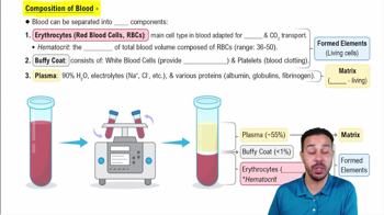
Composition of Blood
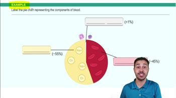
Blood is connective tissue with plasma (nonliving fluid matrix) and formed elements (RBCs, WBCs, platelets).
Physical Characteristics of Blood
Sticky, opaque fluid with metallic taste; color varies with O2 content; pH 7.35–7.45; ~8% body weight; volume 5–6 L males, 4–5 L females.
Hematocrit
Percentage of blood volume occupied by RBCs; normal ~47% males, ~42% females.
Plasma Composition
About 90% water with dissolved solutes: nutrients, gases, hormones, wastes, and plasma proteins like albumin, globulins, and fibrinogen.
Erythrocyte Structure
Biconcave, anucleate cells filled with hemoglobin; flexible due to spectrin; 7.5 μm diameter.
Hemoglobin Structure
Protein globin with four polypeptide chains (2 alpha, 2 beta) each bound to a heme group containing iron that binds O2.
Erythropoiesis
Process of RBC formation in red bone marrow from hematopoietic stem cells through stages including proerythroblast and reticulocyte.
Regulation of Erythropoiesis
Controlled by erythropoietin (EPO) from kidneys in response to hypoxia; requires iron, vitamin B12, and folic acid.
Life Span and Fate of RBCs
Live 100–120 days; old RBCs broken down by spleen macrophages; iron recycled; heme degraded to bilirubin.
Anemia
Condition with low O2-carrying capacity; causes include blood loss, low RBC production, or high RBC destruction.
Sickle-Cell Anemia
Genetic disorder with mutated hemoglobin S causing crescent-shaped RBCs that block vessels and rupture easily.
Polycythemia
Excess RBCs increasing blood viscosity; primary caused by bone marrow cancer, secondary by hypoxia or EPO increase.
Leukocytes (WBCs)
Complete cells with nuclei; defend against disease; classified as granulocytes (neutrophils, eosinophils, basophils) and agranulocytes (lymphocytes, monocytes).
Neutrophils
Most numerous WBCs; phagocytic bacteria slayers; have multilobed nucleus and granules with enzymes and defensins.
Eosinophils
2–4% of WBCs; bilobed nucleus; granules contain enzymes to digest parasitic worms; involved in allergies.
Basophils
Rarest WBCs; granules contain histamine which promotes inflammation and attracts other WBCs.
Lymphocytes
25% of WBCs; large spherical nucleus; key in immunity; T cells attack infected cells, B cells produce antibodies.
Monocytes
Largest WBCs; kidney-shaped nucleus; differentiate into macrophages; phagocytic and activate lymphocytes.
Platelets
Cell fragments from megakaryocytes; involved in clotting by forming platelet plugs and releasing clotting chemicals.
 뒤로
뒤로
 2:36
2:36