 뒤로
뒤로Cardiovascular System and Heart Physiology - Anatomy & Physiology
 6:13
6:13
이 집합의 용어 (27)
The cardiovascular system transports nutrients, gases, and wastes; regulates body temperature and pH; and protects against blood loss and infection.
Blood flows from the aorta to arteries, arterioles, capillaries, venules, veins, and returns to the heart via the venae cavae, completing the circuit back to the aorta.
Blood flows from areas of higher pressure to lower pressure; flow rate depends on pressure gradients, resistance, and vessel radius.
Hydrostatic pressure is the force exerted by blood against vessel walls, driving fluid out of capillaries.
Resistance is inversely proportional to the fourth power of the radius; small changes in radius greatly affect flow and resistance.
The heart has four chambers: two atria and two ventricles, separated by valves that ensure unidirectional blood flow.
The heart is enclosed by the pericardium and has major vessels including the aorta, pulmonary arteries, and veins attached externally.
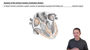
Autorhythmic cells generate electrical impulses; contractile cells produce force for heart contractions.
Myocardial cells are arranged in layers with contractile cells forming the bulk of the myocardium and autorhythmic cells forming the conduction system.
Voltage-gated sodium, calcium, and potassium channels regulate ion movement triggering contraction and relaxation in myocardial cells.
Depolarization opens sodium channels; calcium influx triggers contraction; potassium efflux repolarizes the cell for relaxation.
Autorhythmic cells have spontaneous depolarization; contractile cells have a stable resting potential and longer action potentials.
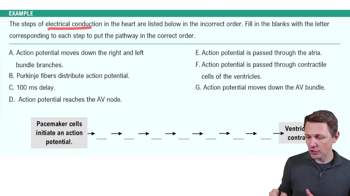
Signals start at the sinoatrial node, pass to the atrioventricular node, then through the bundle of His, bundle branches, and Purkinje fibers.
The ECG includes the P wave (atrial depolarization), QRS complex (ventricular depolarization), and T wave (ventricular repolarization).
Electrical depolarization triggers atrial and ventricular contraction; repolarization corresponds to relaxation phases.
Pressure rises during ventricular systole to eject blood and falls during diastole to allow filling.
Cardiac output = heart rate × stroke volume; it measures blood volume pumped per minute.
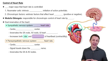
Sympathetic stimulation increases heart rate and contractility; parasympathetic stimulation decreases heart rate.
Stroke volume is affected by venous return, preload, afterload, contractility, skeletal muscle pump, respiratory pump, and inotropic agents.
Length-Tension
Stretching heart muscles optimizes overlap of actin and myosin, producing stronger contraction and larger stroke volume
Afterload
Resistance or blood pressure in arteries and ventricles push to eject blood; increase in it decreases stroke volume
Contractility
Mechanical strength of heart contraction is independent on preload or afterload increase causes increase in stroke volume
Inotropic Agents
Chemical substances/drugs (epinephrine) after contractility; positive increases stroke volume, negative decreases
Venous Return
Increase causes increase in filling ventricles, raising end diastole volume and increasing preload
Preload
Degree of strength on ventricles at the end of diastole increasing causes increases in stroke volume
Skeletal Muscle Pump
Contracting leg and body squeeze veins, pushing blood towards heart and increasing venous and stroke volume
Respiratory Pump
Breathing changes pressure in chest; inhalation lowers pressing, pulling blood towards the heart and enhancing venous