 뒤로
뒤로Cardiovascular System: Blood Vessels, Blood Pressure, and Blood Flow Control
 3:38
3:38
이 집합의 용어 (20)
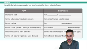
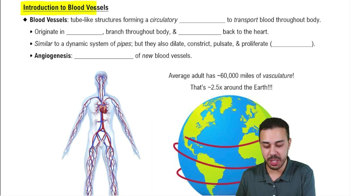
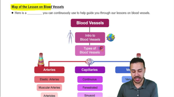
Arteries, arterioles, capillaries, venules, and veins differ in structure, mechanical properties, and function. Arteries have thick muscular walls for high pressure; veins have valves and thinner walls; capillaries allow exchange; arterioles regulate flow; venules collect blood from capillaries.
Blood pressure is highest in the arteries, decreases in arterioles, drops significantly in capillaries, and is lowest in veins as blood returns to the heart.
A cuff inflates to occlude the artery; pressure is slowly released while listening for Korotkoff sounds. The first sound indicates systolic pressure, and disappearance indicates diastolic pressure.
Blood pressure depends on cardiac output (volume pumped by heart) and peripheral resistance (resistance of vessels). Higher output or resistance increases pressure.
Mean arterial pressure is calculated as \(MAP = DP + \frac{1}{3}(SP - DP)\), where SP is systolic pressure and DP is diastolic pressure.
Increased blood volume raises blood pressure by increasing venous return and cardiac output; decreased volume lowers pressure.
Myogenic autoregulation is the intrinsic ability of vascular smooth muscle to respond to stretch by contracting, helping maintain constant blood flow despite pressure changes.
Key paracrine signals include nitric oxide (NO) for vasodilation, endothelin for vasoconstriction, adenosine, and prostaglandins.
Hormones like epinephrine, angiotensin II, and vasopressin regulate vessel diameter by causing vasoconstriction or vasodilation.
Sympathetic nerves release norepinephrine acting on alpha-adrenergic receptors to constrict vessels; beta-adrenergic receptors mediate dilation in some vessels.
Local signaling uses paracrine factors to adjust flow in tissues; long-distance signaling involves nervous and hormonal systems to direct blood flow to or away from organs.
Brain blood flow is tightly regulated by autoregulation via myogenic and metabolic mechanisms to maintain constant perfusion despite systemic pressure changes.
Coronary blood flow increases with heart activity, regulated by local metabolites like adenosine and neural inputs.
Stimulus: increased BP stretches baroreceptors; Sensor: carotid sinus and aortic arch; Input: sensory nerves to medulla; Integrating center: cardiovascular center; Output: autonomic nerves; Targets: heart and vessels; Response: decreased HR and vasodilation; Feedback lowers BP.
Continuous (muscle, brain), fenestrated (kidney, intestines), and sinusoidal (liver, bone marrow) capillaries differ in permeability and structure.
Diffusion moves gases and small solutes across capillary walls; transcytosis transports larger molecules via vesicles.
Hydrostatic pressure pushes fluid out; oncotic pressure pulls fluid in; balance determines net fluid movement.
Lymphatics collect excess interstitial fluid, return it to circulation, and participate in immune defense by transporting lymph and immune cells.
Lymphatics connect to veins, maintain fluid balance, and provide a pathway for immune surveillance and response.
Edema can result from increased capillary hydrostatic pressure, decreased plasma oncotic pressure, lymphatic obstruction, or increased capillary permeability.