 뒤로
뒤로Cardiovascular System II: Blood Vessels - Anatomy & Physiology
 3:38
3:38
이 집합의 용어 (28)
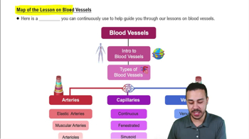
Include arteries, arterioles, capillaries, venules, and veins, each with distinct structure and function.
Heart → arteries → arterioles → capillaries → venules → veins → heart.
Tunica intima (inner), tunica media (middle, smooth muscle), and tunica externa (outer connective tissue).
Controls vasoconstriction (narrowing) and vasodilation (widening) via smooth muscle in tunica media.
Elastic arteries have more elastic fibers for pressure buffering; muscular arteries have more smooth muscle for distribution; arterioles regulate flow to capillaries.
Exchange of materials occurs through capillaries.
Continuous (least permeable), fenestrated (pores for filtration), and sinusoidal (large gaps for cells).
Venules collect blood from capillaries; veins return blood to the heart and have valves.
Prevent backflow of blood and assist venous return to the heart.
Systolic BP is pressure during heart contraction; diastolic BP is pressure during heart relaxation.
Measured in the brachial artery using a sphygmomanometer.
Hypertension is high blood pressure; hypotension is low blood pressure.
Average pressure in arteries during one cardiac cycle; important for tissue perfusion.
Resistance depends on vessel diameter (inversely proportional) and length (directly proportional).
Diameter decreases from elastic arteries to arterioles, then increases in veins.
Viscosity increases resistance; turbulence disrupts flow and increases resistance.
Velocity decreases as total cross-sectional area increases, e.g., slowest in capillaries.
Pulse is the pressure wave caused by heartbeats, felt in arteries.
They aid venous return by compressing veins during breathing and movement.
Vasomotor center controls vessel diameter; cardiac center regulates heart rate and force.
Located in carotid sinuses and aortic arch; detect BP changes and trigger reflexes to maintain homeostasis.
Increased BP stimulates baroreceptors to lower heart rate and dilate vessels; decreased BP has opposite effect.
Located near baroreceptors; detect blood O2, CO2, pH; activation generally increases BP.
Epinephrine/norepinephrine increase BP; aldosterone increases blood volume; antidiuretic hormone promotes water retention; atrial natriuretic peptide lowers BP.
Molecules move down their concentration gradient across capillary walls.
Hydrostatic pressure pushes fluid out of capillaries; colloid osmotic pressure pulls fluid in.
Filtration moves fluid out at arteriole end; reabsorption moves fluid in at venule end of capillaries.
Increased filtration, decreased reabsorption, or lymphatic obstruction can cause fluid accumulation (edema).