 뒤로
뒤로Central Nervous System Anatomy & Physiology
 0:50
0:50
이 집합의 용어 (29)
The CNS consists of the brain and spinal cord, responsible for processing and integrating information.
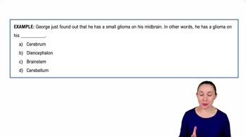
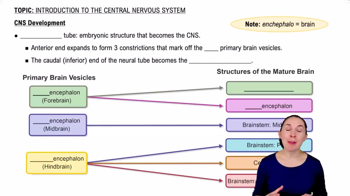
During embryonic development, the brain forms three primary vesicles: prosencephalon (forebrain), mesencephalon (midbrain), and rhombencephalon (hindbrain).
The primary vesicles further divide into five secondary vesicles: telencephalon, diencephalon, mesencephalon, metencephalon, and myelencephalon.
Telencephalon forms cerebral hemispheres; diencephalon forms thalamus and hypothalamus; mesencephalon remains midbrain; metencephalon forms pons and cerebellum; myelencephalon forms medulla oblongata.
Two major flexures at week 5 cause the telencephalon and diencephalon to angle toward the brain stem, shaping brain structure.
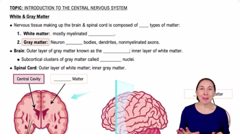
Gray matter consists of neuron cell bodies and short nonmyelinated neurons.
White matter is mostly myelinated axons with some nonmyelinated axons, responsible for communication between brain regions.
In the spinal cord, gray matter is central and white matter is peripheral; in the brain, gray matter forms the cortex and nuclei, with white matter inside.
Four fluid-filled chambers: paired lateral ventricles, third ventricle, and fourth ventricle, all continuous and filled with cerebrospinal fluid.
Make up 83% of brain mass; surface marked by gyri (ridges), sulci (shallow grooves), and fissures (deep grooves).
Longitudinal fissure separates hemispheres; transverse cerebral fissure separates cerebrum and cerebellum.
Five lobes: frontal, parietal, temporal, occipital, and insula (buried under other lobes).
Separates the precentral gyrus (frontal lobe, motor cortex) from the postcentral gyrus (parietal lobe, somatosensory cortex).
Cerebral cortex (gray matter), white matter internally, and basal nuclei deep within white matter.
Thin superficial layer of gray matter containing neuron cell bodies, dendrites, glial cells, and blood vessels; no axons.
Includes motor areas (voluntary movement), sensory areas (conscious sensation), and association areas (integrate information).
Located in the precentral gyrus of the frontal lobe; controls voluntary movements via pyramidal cells.
Plans movements and controls learned, repetitious motor skills; located anterior to primary motor cortex.
Usually in left hemisphere; involved in speech production and planning motor activities for speech.
Located in postcentral gyrus of parietal lobe; receives sensory input from skin and proprioceptors; capable of spatial discrimination.
Posterior to primary somatosensory cortex; integrates sensory input to understand objects by touch.
Primary visual cortex in occipital lobe receives input from retinas; visual association area interprets stimuli using past experience.
Primary auditory cortex in temporal lobe interprets sound; auditory association area stores sound memories and perception.
Located in posterior insula and adjacent parietal cortex; responsible for conscious awareness of balance and head position.
Primary olfactory cortex in temporal lobe (piriform lobes); involved in conscious awareness of odors.
Located in insula; involved in perception of taste.
Posterior to gustatory cortex; conscious perception of visceral sensations like upset stomach or full bladder.
Receive input from multiple sensory areas and send outputs to multiple areas; include anterior, posterior, and limbic association areas.
Division of labor between hemispheres; left controls language, math, logic; right controls visual-spatial skills, intuition, emotion, and artistic skills.