 뒤로
뒤로Central Nervous System Anatomy & Physiology
 0:50
0:50
이 집합의 용어 (24)
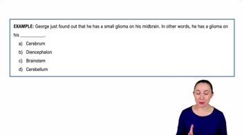
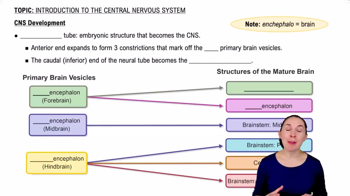
Cerebrum, diencephalon, brainstem, and cerebellum.
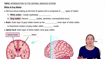
White matter contains myelinated axons; gray matter contains neuron cell bodies and dendrites.
Frontal, parietal, temporal, occipital, and insula lobes with sensory, motor, and association functions.
Sulcus is a shallow groove, gyrus is a ridge, and fissure is a deep groove in the brain.
Association fibers connect areas within the same hemisphere, commissural fibers connect hemispheres, projection fibers connect cortex to lower CNS.
Clusters of gray matter involved in motor control, regulating voluntary movements and posture.
Thalamus (sensory relay), hypothalamus (homeostasis), and epithalamus (pineal gland, circadian rhythms).
Reduced light exposure affects the pineal gland and melatonin production, disrupting mood regulation.
Midbrain, pons, and medulla oblongata, controlling vital functions and pathways.
Coordinates balance, posture, and fine motor control.
Loss of muscle coordination due to cerebellar dysfunction.
Includes hippocampus, amygdala, and others; responsible for emotion and memory.
Located in brainstem; the reticular activating system (RAS) regulates arousal and consciousness.
Broca's area (frontal lobe) controls speech production; Wernicke's area (temporal lobe) controls language comprehension.
Protect CNS; layers are dura mater (outer), arachnoid mater (middle), and pia mater (inner).
Epidural space is outside dura mater, subdural space is between dura and arachnoid, arachnoid space contains cerebrospinal fluid.
CSF cushions CNS, formed by choroid plexus, circulates in ventricles, subarachnoid space, and central canal.
Excess accumulation of CSF causing increased pressure in the brain.
Two lateral ventricles, third ventricle, and fourth ventricle connected by interventricular foramina and cerebral aqueduct.
Selective barrier formed by endothelial cells and astrocytes; protects brain from harmful substances.
Begins at the foramen magnum and ends near L1-L2 vertebrae at the conus medullaris.
Includes cervical and lumbar enlargements, conus medullaris, cauda equina, and filum terminale.
Gray matter forms an H-shape with anterior, posterior, and lateral horns; surrounded by white matter.
Posterior horn contains sensory neurons; anterior horn contains somatic motor neurons; lateral horn contains autonomic motor neurons.