뒤로

Lab 1: Blood

컨트롤 버튼이 '내비게이션' 모드로 변경되었습니다.
1/64
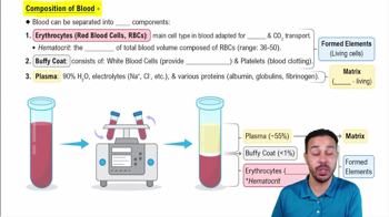
  • What is hematocrit?

    The percentage of blood volume made up of packed red blood cells.
  • What does a high hematocrit mean?

    There are too many red blood cells.
  • What does a low hematocrit mean?

    There are not enough red blood cells.
  • What is the normal hematocrit range?

    Adult males: 47% ± 5%. Adult females: 42% ± 5%.
  • What is the normal pH range of blood?

    7.35–7.45.
  • What is plasma?

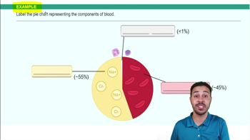
    The liquid extracellular portion of blood that the blood cells and cell fragments are suspended in.
  • What are the formed elements of blood?

    Erythrocytes, leukocytes, and platelets.
  • What is another name for an erythrocyte?

    Red blood cell (RBC).
  • What do erythrocytes do?

    Carry oxygen and carbon dioxide.
  • Do mature erythrocytes have a nucleus?

    No. They are anucleate.
  • What does an erythrocyte look like under the microscope?

    A pink/red biconcave disc with a pale center and no nucleus.
  • What is another name for a leukocyte?

    White blood cell (WBC).
  • What is the main function of leukocytes?

    They provide immunity and help protect the body.
  • Do leukocytes have a nucleus?

    Yes. Leukocytes are nucleated cells.
  • What are the 5 types of leukocytes?

    Neutrophils, eosinophils, basophils, lymphocytes, and monocytes.
  • What are the 3 granulocytes?

    Neutrophils, eosinophils, and basophils.
  • What are the 2 agranulocytes?

    Lymphocytes and monocytes.
  • What does Never Let Monkeys Eat Bananas stand for?


    Neutrophils, Lymphocytes, Monocytes, Eosinophils, Basophils

  • What is a neutrophil?

    A granulocyte that fights bacteria and is the most common circulating WBC.
  • What percentage of circulating WBCs are neutrophils?

    About 50–70%.
  • How do you recognize a neutrophil under the microscope?

    Multilobed nucleus, usually 2–5 lobes, with pale/light purple granules.
  • What is the function of an eosinophil?

    Helps fight parasites and is involved in allergies and asthma.
  • How do you recognize an eosinophil under the microscope?

    Bilobed nucleus with reddish/orange cytoplasmic granules.
  • What is the function of a basophil?

    Involved in inflammation and releases histamine and heparin.
  • How do you recognize a basophil under the microscope?

    Dark purple granules that can make the nucleus difficult to see.
  • What is the function of lymphocytes?

    T and B lymphocytes help with immunity. B cells make antibodies, and lymphocytes help fight viruses and cancer cells.
  • How do you recognize a lymphocyte under the microscope?

    Large, round/spherical nucleus that takes up most of the cell with only a thin rim of cytoplasm.
  • What is a monocyte?

    A large agranulocyte associated with chronic infections that can develop into a macrophage in tissues.
  • How do you recognize a monocyte under the microscope?

    Large cell with a kidney-shaped or U-shaped nucleus and abundant pale cytoplasm.
  • How can you remember the 5 WBCs?

    Never Let Monkeys Eat Bananas = Neutrophils, Lymphocytes, Monocytes, Eosinophils, Basophils.
  • What is a differential white blood cell count?

    A test that determines the relative percentage of each type of white blood cell.
  • Why is a differential WBC count performed?

    It can help detect conditions such as acute infection, chronic infection, allergies, parasitic disease, anemia, HIV infection, and others.
  • What is another name for a platelet?

    Thrombocyte.
  • What are platelets?

    Small cell fragments involved in hemostasis.
  • What is the main function of platelets?

    Help stop bleeding through hemostasis.
  • How do you recognize platelets under the microscope?

    Very small, irregular cell fragments scattered between the much larger red blood cells.
  • What is hemostasis?

    The process of stopping bleeding.
  • What does the word hemostasis mean?

    “Hemo” = blood and “stasis” = stop.
  • What stain is used on prepared blood smears in Lab 1?

    Wright's stain.
  • What dyes are found in Wright's stain?

    Methylene blue and eosin diluted in methanol.
  • What does eosinophilic material look like after Wright's staining?

    Pink, red, or orange-red.
  • What does basophilic material look like after Wright's staining?

    Blue to dark purple.
  • What is agglutination?

    Clumping of red blood cells caused by an antigen-antibody reaction.
  • How is agglutination used in blood typing?

    Clumping shows that the antigen being tested for is present on the red blood cells.
  • What does the D antigen determine?

    The Rh factor.
  • What does it mean if the D antigen is present?

    The blood type is Rh positive (+).
  • What does it mean if the D antigen is absent?

    The blood type is Rh negative (−).
  • If blood clumps with Anti-A, what antigen is present?

    A antigen.
  • If blood clumps with Anti-B, what antigen is present?

    B antigen.
  • If blood clumps with Anti-D, what does that mean?

    The person is Rh positive.
  • Anti-A clumps only = what blood type?

    Type A.
  • Anti-B clumps only = what blood type?

    Type B.
  • Anti-A AND Anti-B both clump = what blood type?

    Type AB.
  • No reaction with Anti-A or Anti-B = what blood type?

    Type O.
  • If Anti-D clumps, what symbol is added to the blood type?

    Positive (+).
  • If Anti-D does NOT clump, what symbol is added?

    Negative (−).
  • Who can an Rh-positive person receive blood from regarding Rh factor?

    Rh-positive or Rh-negative blood.
  • Who can an Rh-positive person donate blood to regarding Rh factor?

    Other Rh-positive people.
  • Who can O receive blood from?


    Only O

  • Who can A receive blood from?


    A and O

  • Who can B receive blood from?


    B and O

  • Who can AB receive blood from?


    AB, A, B, and O

  • Who can Rh+ receive blood from?


    Rh+ and Rh-

  • Who can Rh- receive blood from?


    Only Rh-