 뒤로
뒤로Lab 3: Blood Vessel;s
 3:38
3:38
이 집합의 용어 (22)
Artery
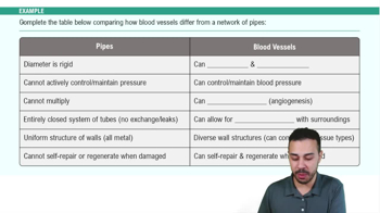
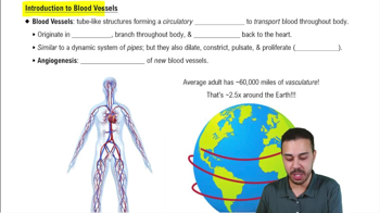
Carries blood AWAY from the heart
Has a thicker wall than a vein
Has a thick tunica media
Usually has a smaller, rounder lumen
Does not have valves
Usually red on the lab model
Thick walls help arteries withstand the higher blood pressure coming from the heart
🧠 A = Artery = Away
Vein
Carries blood TOWARD the heart
Thinner wall than an artery
Thinner tunica media
Usually has a larger lumen
Can contain valves that prevent backflow
Usually blue on the lab model
Veins drain blood from tissues and return it toward the heart
🧠 Veins = back TO the heart
Capillary
Smallest type of blood vessel
Located between the arterial and venous sides of circulation
Forms capillary beds
Main location for exchange of O₂, CO₂, nutrients, and wastes
Very thin wall allows substances to move between blood and tissues
🧠 Your note: Capillary beds = where exchange happens.
Tunica Intima (Interna)
Innermost layer of a blood vessel wall
Directly contacts the blood
Contains endothelium
🧠 Intima = IN
Tunica Media
Middle layer of the blood vessel wall
Contains mainly smooth muscle
Thick in arteries
Thin in veins
⭐ Thick tunica media on histology = think ARTERY
Tunica Externa (Adventitia)
Outermost layer of a blood vessel wall
Made mainly of connective tissue
Supports and protects the vessel
🧠 Externa = exterior/outside
Lumen
Hollow space inside a blood vessel where blood flows
Arteries usually have a smaller lumen
Veins usually have a larger lumen
🧠 Small lumen + thick wall = artery
Aorta
Largest artery in the body
Begins at the left ventricle
Main artery of systemic circulation
Gives rise to arteries that supply the body
🧠 Model: Major red vessel leaving the left side of the heart.
Ascending Aorta
First portion of the aorta
Location: Rises upward from the left ventricle
Leads into the aortic arch
Coronary arteries branch from this region
🧠 Ascending = going UP
Aorta Arch
Curved portion of the aorta
Location: Superior to the heart
Between the ascending and descending aorta
Gives off major branches supplying the head, neck, and upper limbs
🧠 Look for the large curve/arch above the heart.
Descending Aorta
Portion of aorta that travels downward
Continues from the aortic arch
Includes the thoracic and abdominal portions
🧠 Descending = going DOWN
Thoracic Aorta
Portion of the descending aorta located in the thoracic/chest cavity
Continues inferiorly and becomes the abdominal aorta
🧠 Thoracic = chest
Brachiocephalic Trunk
First branch of the aortic arch
Located on the right side
Divides into:
Right common carotid artery
Right subclavian artery
⭐ There is only ONE brachiocephalic arterial trunk.
🧠 Common = think split
R/L Common Carotid Arteries
Location: Travel superiorly through the neck
Supply the head and neck
Each divides into:
Internal carotid
External carotid
🧠 Common carotid → splits into internal + external
R/L External Carotid Arteries
Branches of the common carotid arteries
Location: Neck/head
Primarily supply structures outside the skull, especially the face and scalp
🧠 External = outside
R/L Internal Carotid Arteries
Branches of the common carotid arteries
Travel toward the inside of the skull
Major blood supply to the brain
🧠 Internal = inside → brain
R/L Subclavian Arteries
Location: Beneath the clavicles
Supply the upper limbs
Continue into the axillary arteries
🧠 Sub = under + clavian = clavicle
R/L Vertebral Arteries
Branch from the subclavian arteries
Travel superiorly through the neck along the vertebral column
Help supply the brain
🧠 Vertebral = vertebrae
Intercostal Arteries
Branches associated with the thoracic aorta
Location: Run between the ribs
Supply the thoracic wall/intercostal spaces
🧠 Intercostal = between ribs
R/L Subscapular Arteries
Branches associated with the axillary artery
Location: Near the scapular/shoulder region
Supply structures around the scapula
🧠 Subscapular = around/below scapula
R/L Coronary Arteries
Branch from the base of the ascending aorta
Located on the surface of the heart
Supply the heart muscle (myocardium) itself
🧠 Coronary = heart's own blood supply
Internal Thoracic (Mammary) Artery
Branch of the subclavian artery
Travels along the inside of the anterior chest wall
Supplies structures of the thoracic/chest wall
🧠 Thoracic = chest