Skip to main content
Anatomy & Physiology
나의 코스
배우기
시험 준비
AI 튜터
학습 가이드
교과서적 해결책
플래시카드
탐험해 보세요
앱을 사용해 보세요
나의 코스
배우기
시험 준비
AI 튜터
학습 가이드
교과서적 해결책
플래시카드
탐험해 보세요
앱을 사용해 보세요
뒤로
Muscle Structure and Contraction - Anatomy & Physiology
카드를 뒤집기 위해 탭할 수 있습니다.
Three types of muscle tissue
카드를 뒤집기 위해 탭할 수 있습니다.
👆
Three types of muscle tissue
Skeletal
,
cardiac
, and
smooth
muscle differ in structure, location, function, and activation.
진행 상황 추적
컨트롤 버튼이 '내비게이션' 모드로 변경되었습니다.
1/21
추천 영상
6:31
Types of Muscle Tissue
15902
views
435
rank
1
comments
2:00
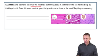
Introduction to Muscles and Muscle Tissue Example 1
13554
views
279
rank
4:36
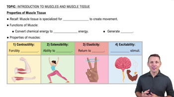
Properties of Muscle Tissue
21903
views
634
rank
2
comments
이 집합의 용어 (21)
하이드의 정의
Three types of muscle tissue
Skeletal
,
cardiac
, and
smooth
muscle differ in structure, location, function, and activation.
Muscle fiber
An elongated muscle cell, also called a muscle fiber, found in skeletal and smooth muscle.
Connective tissue sheaths in skeletal muscle
Endomysium
surrounds each muscle fiber,
perimysium
surrounds fascicles, and
epimysium
surrounds the entire muscle.
Sarcomere
The smallest contractile unit of muscle, between two Z discs, composed of thick (myosin) and thin (actin) filaments.
Thick filaments composition
Composed of
myosin
molecules, each with a rodlike tail and two globular heads that form cross bridges.
Thin filaments composition
Primarily composed of
actin
with regulatory proteins
tropomyosin
and
troponin
bound to actin.
Role of calcium in muscle contraction
Calcium binds to troponin, causing tropomyosin to move and expose myosin binding sites on actin, enabling contraction.
Sarcoplasmic reticulum (SR)
A smooth ER that stores and regulates intracellular calcium, surrounding each myofibril and forming terminal cisternae.
T tubules function
Invaginations of the sarcolemma that conduct action potentials deep into muscle fibers, triggering Ca2+ release from SR.
Sliding filament model
Thin filaments slide past thick filaments, shortening sarcomeres and causing muscle contraction.
Neuromuscular junction
Synapse between a motor neuron axon terminal and a muscle fiber's motor end plate where acetylcholine is released.
Acetylcholine (ACh) role
Binds to receptors on the sarcolemma, opening Na+ channels, causing depolarization and initiating muscle action potential.
Excitation-contraction coupling
Process linking muscle action potential to contraction via Ca2+ release from SR and exposure of actin binding sites.
Cross bridge cycle
Myosin heads attach to actin, pivot to pull thin filaments, detach with ATP binding, and reset for another cycle.
Motor unit
A motor neuron and all the muscle fibers it innervates; size varies with muscle function.
Muscle twitch phases
Latent period (excitation-contraction coupling), contraction period (cross bridge activity), and relaxation period (Ca2+ reuptake).
Graded muscle responses
Variations in contraction strength achieved by changing stimulus frequency or strength (recruitment).
Isotonic vs isometric contraction
Isotonic
: muscle changes length and moves load;
Isometric
: muscle tension increases without length change.
Sources of ATP for muscle contraction
ATP is regenerated by creatine phosphate, anaerobic glycolysis, and aerobic respiration.
Muscle fatigue causes
ATP deficit, lactic acid buildup, and ionic imbalances leading to inability to contract.
Muscle tone
Constant, slight contraction of muscles maintained by spinal reflexes to keep muscles firm and ready.
 뒤로뒤로
뒤로뒤로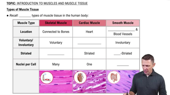
 6:31
6:31