Skip to main content
Anatomy & Physiology
나의 코스
배우기
시험 준비
AI 튜터
학습 가이드
교과서적 해결책
플래시카드
탐험해 보세요
앱을 사용해 보세요
나의 코스
배우기
시험 준비
AI 튜터
학습 가이드
교과서적 해결책
플래시카드
탐험해 보세요
앱을 사용해 보세요
뒤로
Muscle Tissue and Contraction - Anatomy & Physiology
카드를 뒤집기 위해 탭할 수 있습니다.
Main function of muscle tissue
카드를 뒤집기 위해 탭할 수 있습니다.
👆
Main function of muscle tissue
Convert chemical energy to
mechanical energy
to create movement and generate
force
.
진행 상황 추적
컨트롤 버튼이 '내비게이션' 모드로 변경되었습니다.
1/22
추천 영상
6:31
Types of Muscle Tissue
15905
views
435
rank
1
comments
2:00
Introduction to Muscles and Muscle Tissue Example 1
13560
views
279
rank
4:36
Properties of Muscle Tissue
21908
views
634
rank
2
comments
이 집합의 용어 (22)
하이드의 정의
Main function of muscle tissue
Convert chemical energy to
mechanical energy
to create movement and generate
force
.
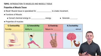
Four properties of muscle tissue
Contractility
: forcibly shorten;
Extensibility
: ability to stretch;
Elasticity
: return to original length;
Excitability
: respond to stimuli.
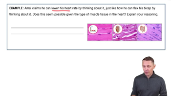
Types of muscle tissue in the human body
Skeletal
,
Cardiac
, and
Smooth
muscle tissues.
Which muscle types are striated?
Skeletal
and
Cardiac
muscles are striated;
Smooth
muscle is non-striated.
Voluntary vs involuntary muscle control
Skeletal muscle
is voluntary;
Cardiac
and
Smooth
muscles are involuntary.
Largest muscle cell type
Skeletal muscle cells
are typically the largest muscle cells.
Organization of skeletal muscle tissue
Muscle fibers bundled into
fascicles
, fascicles bundled into
muscle
, surrounded by connective tissues:
endomysium
,
perimysium
, and
epimysium
.
Connective tissue connections to bones
Muscles connect to bones via
tendon-like
and
aponeurosis-like
structures.
Structure of a muscle fiber
A long, multinucleated cell surrounded by the
endomysium
and plasma membrane called the
sarcolemma
.
What is a sarcomere?
The
smallest contractile unit
of a muscle fiber, composed of actin and myosin filaments.
Sliding filament theory
Muscle shortens as
myosin
(thick filament) pulls on
actin
(thin filament), increasing overlap without changing filament length.
Contractile proteins of the sarcomere
Myosin
(thick filament) and
Actin
(thin filament).
Regulatory proteins in muscle contraction
Tropomyosin
blocks myosin binding sites on actin;
Troponin
binds Ca+2 and moves tropomyosin to expose binding sites.
Structural protein in sarcomere
Titin
provides elasticity and helps sarcomere return to resting length.
Regions of the sarcomere
Z disc
: sarcomere boundary;
M line
: middle anchoring myosin;
A band
: length of myosin;
I band
: actin only;
H zone
: myosin only.
What shortens during muscle contraction?
The
sarcomere
shortens; the
I band
and
H zone
decrease;
A band
length remains constant.
Role of the sarcoplasmic reticulum in contraction
Stores and releases
Ca+2 ions
to trigger muscle contraction.
Excitation-contraction coupling
Process where an
action potential
triggers Ca+2 release, exposing myosin binding sites and causing contraction.
Neurotransmitter at neuromuscular junction
Acetylcholine (ACh)
is released to stimulate muscle fiber contraction.
Phases of action potential in muscle fiber
Depolarization
: Na+ enters cell making inside positive;
Repolarization
: K+ exits restoring negative charge.
Cross-bridge cycle steps
1) Myosin binds actin; 2) Power stroke pulls actin; 3) ATP binds myosin releasing actin; 4) ATP hydrolysis cocks myosin head.
Effect of ATP depletion on muscle contraction
Myosin remains bound to actin causing muscle rigidity; contraction cannot continue.
 뒤로뒤로
뒤로뒤로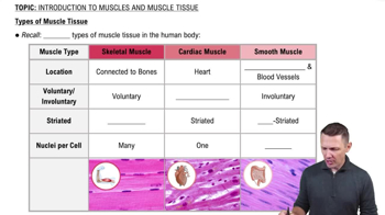
 6:31
6:31